- Neurosurgical and Vascular Surgical anatomy

Содержание
- 2. Outline Sectional imaging of the brain Anatomy Detecting pathology Common vascular surgery operations
- 3. Neurosurgical anatomy
- 4. Sectional anatomy :Brain Cerebrum Cerebellum Brainstem
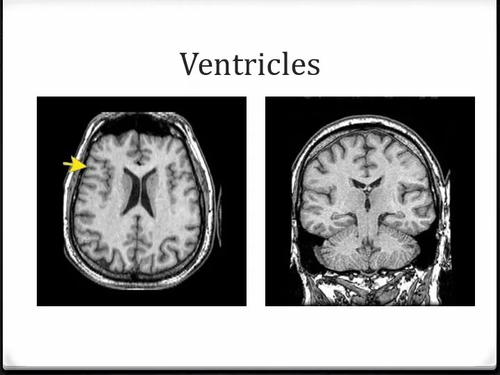
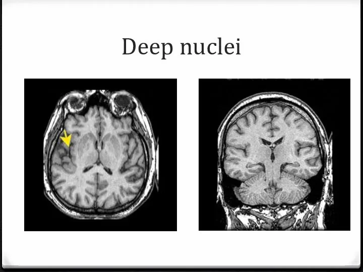
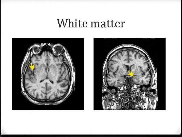
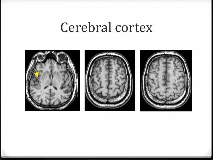
- 5. Cerebrum 4 layers Ventricles Deep nuclei White matter Cortex
- 6. Ventricles
- 7. Deep nuclei
- 8. White matter
- 9. Cerebral cortex
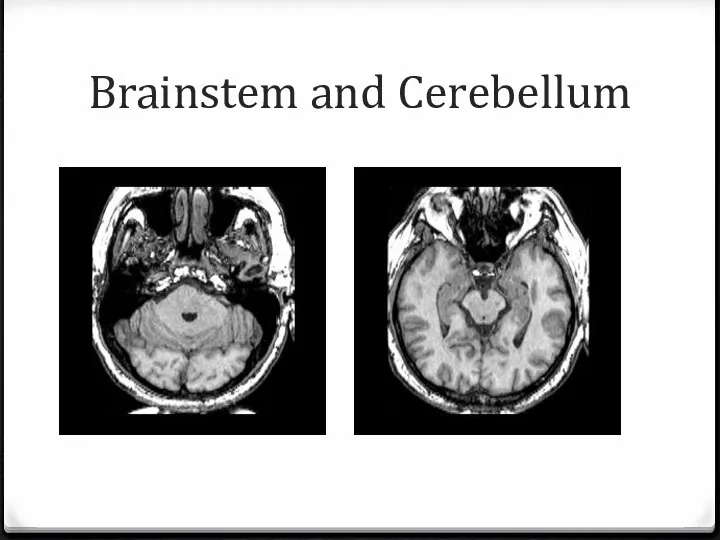
- 10. Brainstem and Cerebellum
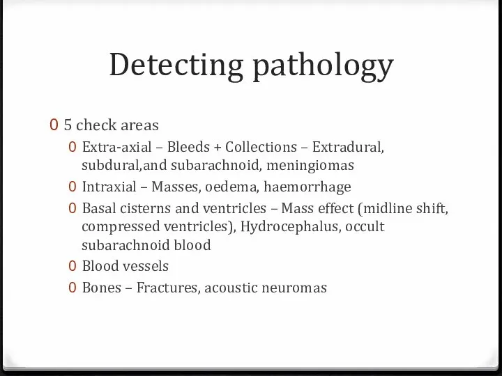
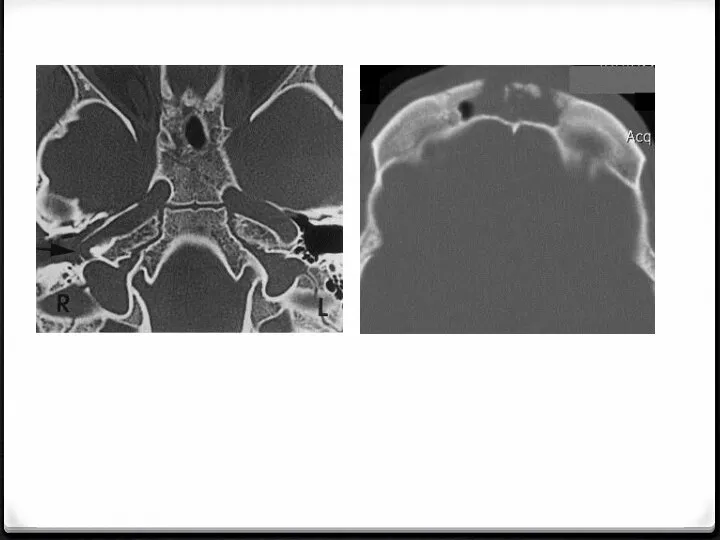
- 11. Detecting pathology 5 check areas Extra-axial – Bleeds + Collections – Extradural, subdural,and subarachnoid, meningiomas Intraxial
- 19. Vascular surgical anatomy
- 20. Common operations Common carotid artery Brachial Radial Femoral Popliteal
- 21. Common carotid a Dangers Layers
- 23. Brachial a Dangers Layers
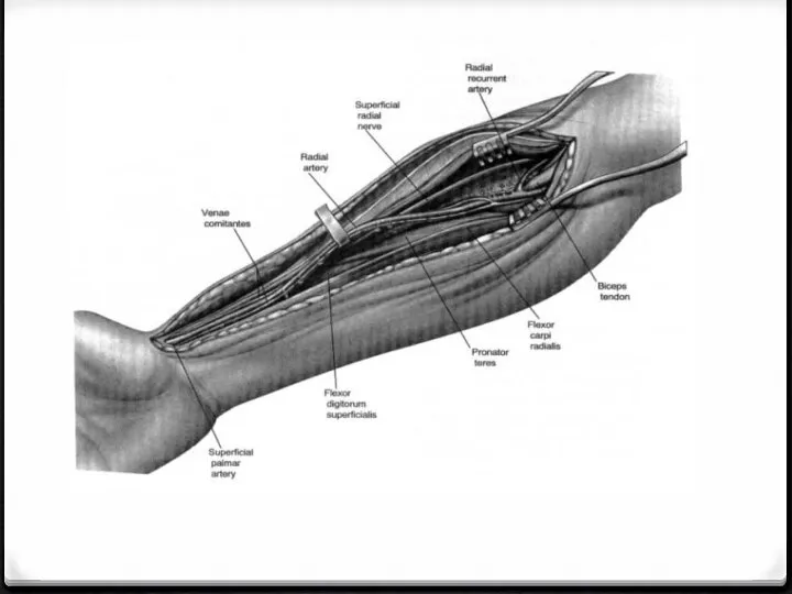
- 25. Radial a Dangers Layers
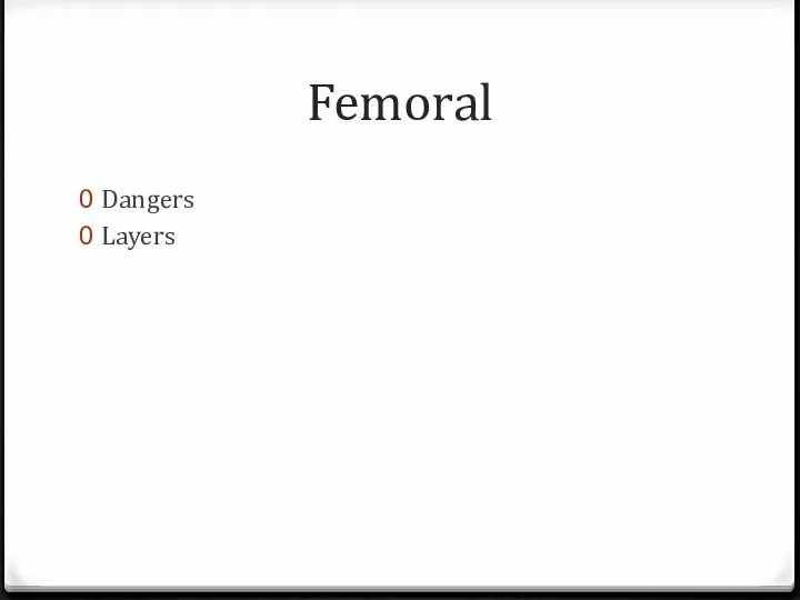
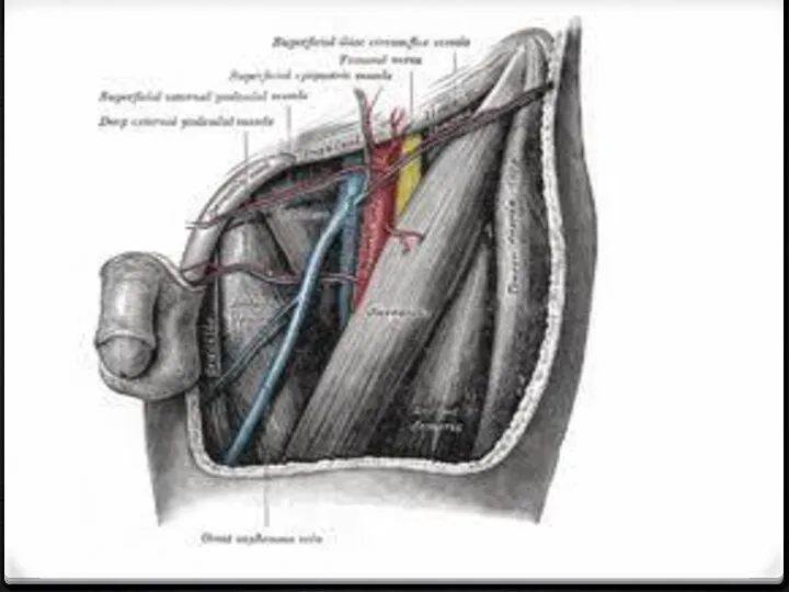
- 27. Femoral Dangers Layers
- 29. Popliteal artery Dangers Layers
- 31. Скачать презентацию
Слайд 2Outline
Sectional imaging of the brain
Anatomy
Detecting pathology
Common vascular surgery operations
Outline
Sectional imaging of the brain
Anatomy
Detecting pathology
Common vascular surgery operations
Слайд 3Neurosurgical anatomy
Neurosurgical anatomy
Слайд 4Sectional anatomy :Brain
Cerebrum
Cerebellum
Brainstem
Sectional anatomy :Brain
Cerebrum
Cerebellum
Brainstem
Слайд 5Cerebrum
4 layers
Ventricles
Deep nuclei
White matter
Cortex
Cerebrum
4 layers
Ventricles
Deep nuclei
White matter
Cortex
Слайд 6Ventricles
Ventricles
Слайд 7Deep nuclei
Deep nuclei
Слайд 8White matter
White matter
Слайд 9Cerebral cortex
Cerebral cortex
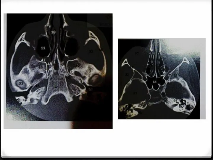
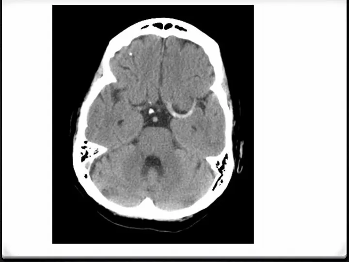
Слайд 10Brainstem and Cerebellum
Brainstem and Cerebellum
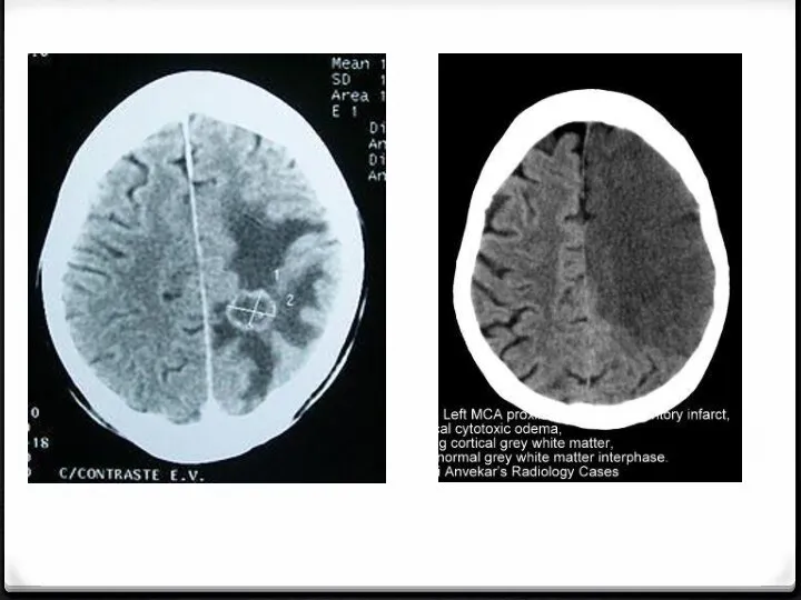
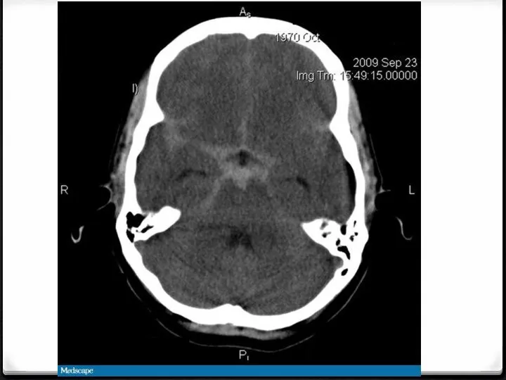
Слайд 11Detecting pathology
5 check areas
Extra-axial – Bleeds + Collections – Extradural, subdural,and subarachnoid,
Detecting pathology
5 check areas
Extra-axial – Bleeds + Collections – Extradural, subdural,and subarachnoid,
Слайд 19Vascular surgical anatomy
Vascular surgical anatomy
Слайд 20Common operations
Common carotid artery
Brachial
Radial
Femoral
Popliteal
Common operations
Common carotid artery
Brachial
Radial
Femoral
Popliteal
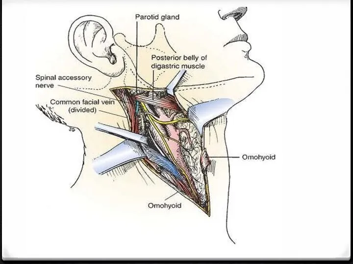
Слайд 21Common carotid a
Dangers
Layers
Common carotid a
Dangers
Layers
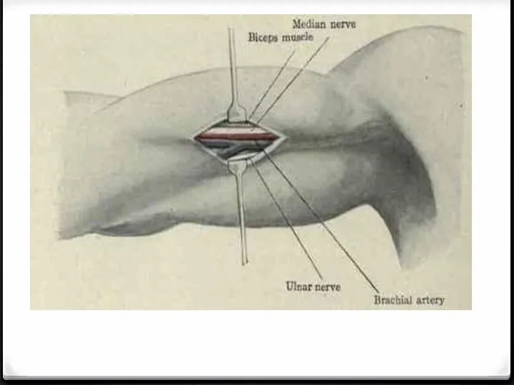
Слайд 23Brachial a
Dangers
Layers
Brachial a
Dangers
Layers
Слайд 25Radial a
Dangers
Layers
Radial a
Dangers
Layers
Слайд 27Femoral
Dangers
Layers
Femoral
Dangers
Layers
Слайд 29Popliteal artery
Dangers
Layers
Popliteal artery
Dangers
Layers
 Амбулаториялық практикада жансыздандыру әдістерін талғау
 Медицинская реабилитация при онкопатологии
 Дизентерийная амёба (лат. Entamoeba histolytica)
 Наркомания
 Дезинфекционно-стерилизационные мероприятия в обеспечении противоэпидемического режима. Обработка рук
 Полезные продукты
 Медицина народного доверия: Служение стране. Служение людям
 Математические модели анализа медицинских данных, методы и алгоритмы их реализации
 Искусство мануальных
 Спируратозы и рабдиатозы
 Лекция 5 ФКТ,цирротический
 Саккодированное и пуэрильное дыхания. Этиология, патогенез, клиника
 Гипертоническая болезнь
 Кишечная пластика мочевого пузыря после цистэктомии,выполненной по поводу рака и его морфо-функциональные последствия к.м.н.,доце
 Хроническая обструктивная болезнь легких. Школа для врачей
 Мочевыделительная система. Кожа
 Методы лечения наследственных патологий. Медико-генетическое консультирование. Тема 11
 Школа аутизма. Особенности развития речевой сферы у детей с аутизмом
 История развития психиатрии XVIII-XIX века: заслуги Ф. Пинеля и Дж. Конолли
 Группы лекарственных средств. Практическая работа
 Нозокомиальные инфекции
 Диабету предшествует предиабет
 Бесплодие. Синдром аменореи
 Клинический случай
 Влияние факторов окружающей среды и социальных условий на здоровье ребенка
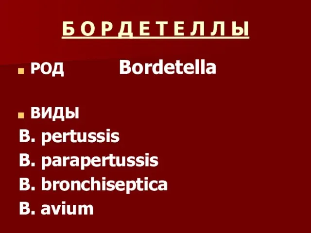 Бордетелла. Виды бордетеллы
 Токсическое действие сердечных гликозидов
 Отравление угарным газом