- SummaryofFunctionofCranial Nerves

Содержание
- 2. Cranial Nerve I: Olfactory Arises from the olfactory epithelium Passes through the cribriform plate of the
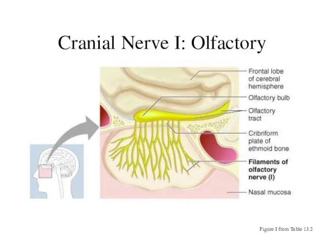
- 3. Cranial Nerve I: Olfactory Figure I from Table 13.2
- 4. Cranial Nerve II: Optic Arises from the retina of the eye Optic nerves pass through the
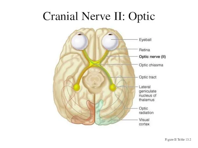
- 5. Cranial Nerve II: Optic Figure II Table 13.2
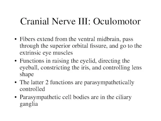
- 6. Cranial Nerve III: Oculomotor Fibers extend from the ventral midbrain, pass through the superior orbital fissure,
- 7. Cranial Nerve III: Oculomotor Figure III from Table 13.2
- 8. Cranial Nerve IV: Trochlear Fibers emerge from the dorsal midbrain and enter the orbits via the
- 9. Cranial Nerve IV: Trochlear Figure IV from Table 13.2
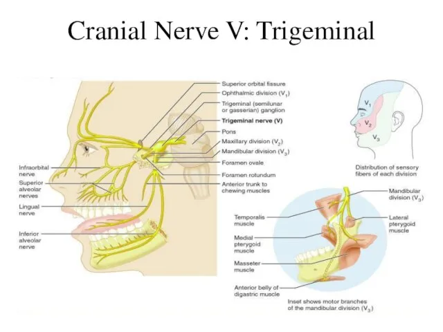
- 10. Cranial Nerve V: Trigeminal Composed of three divisions Ophthalmic (V1) Maxillary (V2) Mandibular (V3) Fibers run
- 11. Cranial Nerve V: Trigeminal
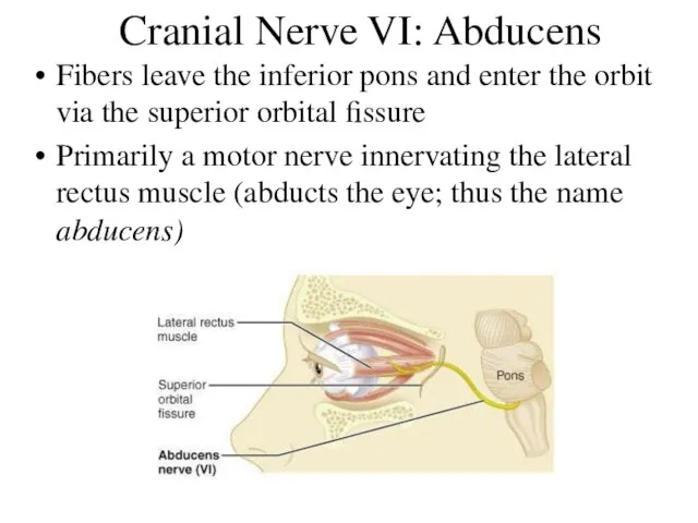
- 12. Cranial Nerve VI: Abducens Fibers leave the inferior pons and enter the orbit via the superior
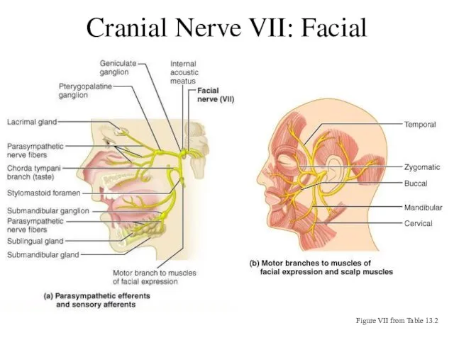
- 13. Cranial Nerve VII: Facial Fibers leave the pons, travel through the internal acoustic meatus, and emerge
- 14. Cranial Nerve VII: Facial Figure VII from Table 13.2
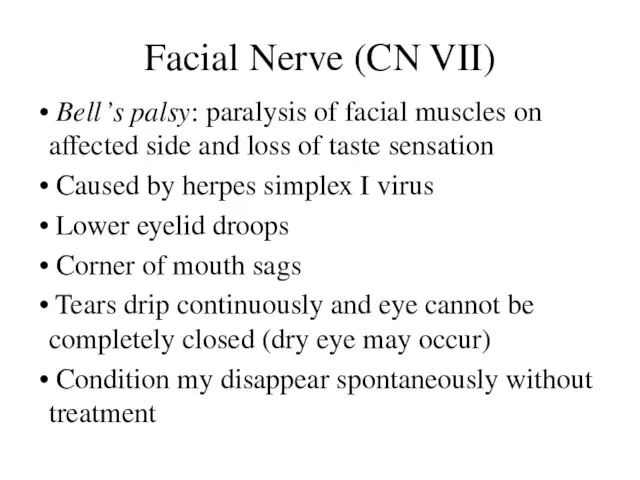
- 15. Facial Nerve (CN VII) Bell’s palsy: paralysis of facial muscles on affected side and loss of
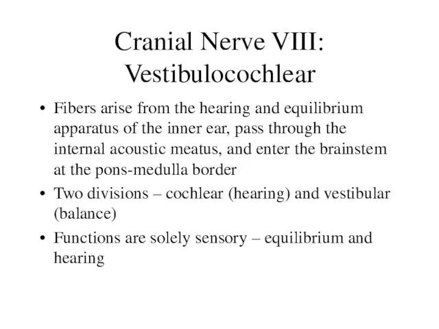
- 16. Cranial Nerve VIII: Vestibulocochlear Fibers arise from the hearing and equilibrium apparatus of the inner ear,
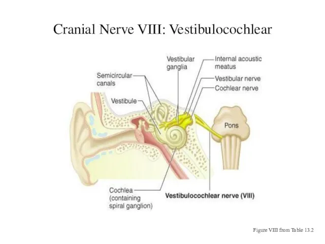
- 17. Cranial Nerve VIII: Vestibulocochlear Figure VIII from Table 13.2
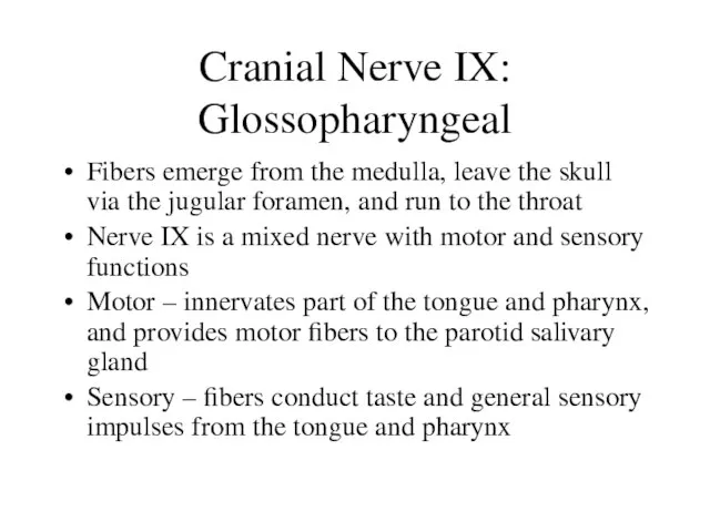
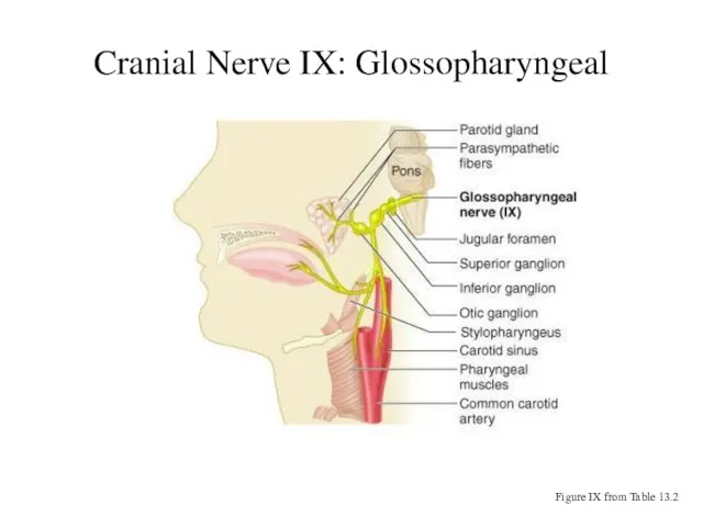
- 18. Cranial Nerve IX: Glossopharyngeal Fibers emerge from the medulla, leave the skull via the jugular foramen,
- 19. Cranial Nerve IX: Glossopharyngeal Figure IX from Table 13.2
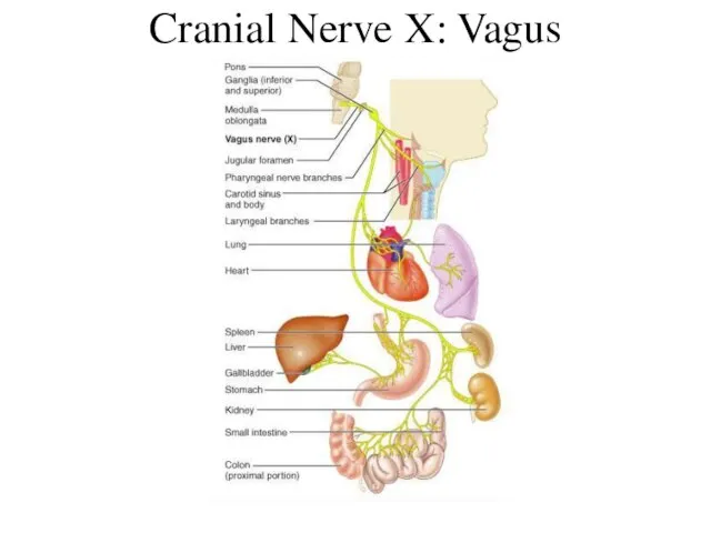
- 20. Cranial Nerve X: Vagus The only cranial nerve that extends beyond the head and neck Fibers
- 21. Cranial Nerve X: Vagus
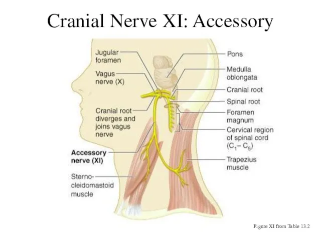
- 22. Cranial Nerve XI: Accessory Formed from a cranial root emerging from the medulla and a spinal
- 23. Cranial Nerve XI: Accessory Figure XI from Table 13.2
- 24. Cranial Nerve XII: Hypoglossal Fibers arise from the medulla and exit the skull via the hypoglossal
- 26. Скачать презентацию
Слайд 2Cranial Nerve I: Olfactory
Arises from the olfactory epithelium
Passes through the cribriform plate
Cranial Nerve I: Olfactory
Arises from the olfactory epithelium
Passes through the cribriform plate
Слайд 3Cranial Nerve I: Olfactory
Figure I from Table 13.2
Cranial Nerve I: Olfactory
Figure I from Table 13.2
Слайд 4Cranial Nerve II: Optic
Arises from the retina of the eye
Optic nerves pass
Cranial Nerve II: Optic
Arises from the retina of the eye
Optic nerves pass
Слайд 5Cranial Nerve II: Optic
Figure II Table 13.2
Cranial Nerve II: Optic
Figure II Table 13.2
Слайд 6Cranial Nerve III: Oculomotor
Fibers extend from the ventral midbrain, pass through the
Cranial Nerve III: Oculomotor
Fibers extend from the ventral midbrain, pass through the
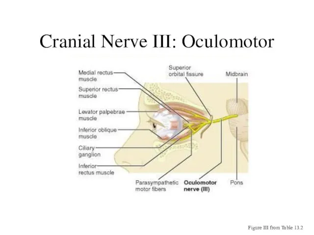
Слайд 7Cranial Nerve III: Oculomotor
Figure III from Table 13.2
Cranial Nerve III: Oculomotor
Figure III from Table 13.2
Слайд 8Cranial Nerve IV: Trochlear
Fibers emerge from the dorsal midbrain and enter the
Cranial Nerve IV: Trochlear
Fibers emerge from the dorsal midbrain and enter the
Слайд 9Cranial Nerve IV: Trochlear
Figure IV from Table 13.2
Cranial Nerve IV: Trochlear
Figure IV from Table 13.2

Слайд 10Cranial Nerve V: Trigeminal
Composed of three divisions
Ophthalmic (V1)
Maxillary (V2)
Mandibular (V3)
Fibers run from
Cranial Nerve V: Trigeminal
Composed of three divisions
Ophthalmic (V1)
Maxillary (V2)
Mandibular (V3)
Fibers run from
Слайд 11Cranial Nerve V: Trigeminal
Cranial Nerve V: Trigeminal
Слайд 12Cranial Nerve VI: Abducens
Fibers leave the inferior pons and enter the orbit
Cranial Nerve VI: Abducens
Fibers leave the inferior pons and enter the orbit
Слайд 13Cranial Nerve VII: Facial
Fibers leave the pons, travel through the internal acoustic
Cranial Nerve VII: Facial
Fibers leave the pons, travel through the internal acoustic
Слайд 14Cranial Nerve VII: Facial
Figure VII from Table 13.2
Cranial Nerve VII: Facial
Figure VII from Table 13.2
Слайд 15Facial Nerve (CN VII)
Bell’s palsy: paralysis of facial muscles on affected
Facial Nerve (CN VII)
Bell’s palsy: paralysis of facial muscles on affected
Слайд 16Cranial Nerve VIII: Vestibulocochlear
Fibers arise from the hearing and equilibrium apparatus of
Cranial Nerve VIII: Vestibulocochlear
Fibers arise from the hearing and equilibrium apparatus of
Слайд 17Cranial Nerve VIII: Vestibulocochlear
Figure VIII from Table 13.2
Cranial Nerve VIII: Vestibulocochlear
Figure VIII from Table 13.2
Слайд 18Cranial Nerve IX: Glossopharyngeal
Fibers emerge from the medulla, leave the skull via
Cranial Nerve IX: Glossopharyngeal
Fibers emerge from the medulla, leave the skull via
Слайд 19Cranial Nerve IX: Glossopharyngeal
Figure IX from Table 13.2
Cranial Nerve IX: Glossopharyngeal
Figure IX from Table 13.2
Слайд 20Cranial Nerve X: Vagus
The only cranial nerve that extends beyond the head
Cranial Nerve X: Vagus
The only cranial nerve that extends beyond the head
Слайд 21Cranial Nerve X: Vagus
Cranial Nerve X: Vagus
Слайд 22Cranial Nerve XI: Accessory
Formed from a cranial root emerging from the medulla
Cranial Nerve XI: Accessory
Formed from a cranial root emerging from the medulla
Слайд 23Cranial Nerve XI: Accessory
Figure XI from Table 13.2
Cranial Nerve XI: Accessory
Figure XI from Table 13.2
Слайд 24Cranial Nerve XII: Hypoglossal
Fibers arise from the medulla and exit the skull
Cranial Nerve XII: Hypoglossal
Fibers arise from the medulla and exit the skull
 Вов
Вов Бальні танці
 Состояние и перспективы развития системы аккредитации в сфере добровольного и обязательного подтверждения соответствия в Герман
 С любовью к животным
 Ооо Невская химия
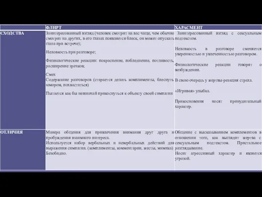 Психология человека
 Практические аспекты диагностики и лечения тромбозов у детей
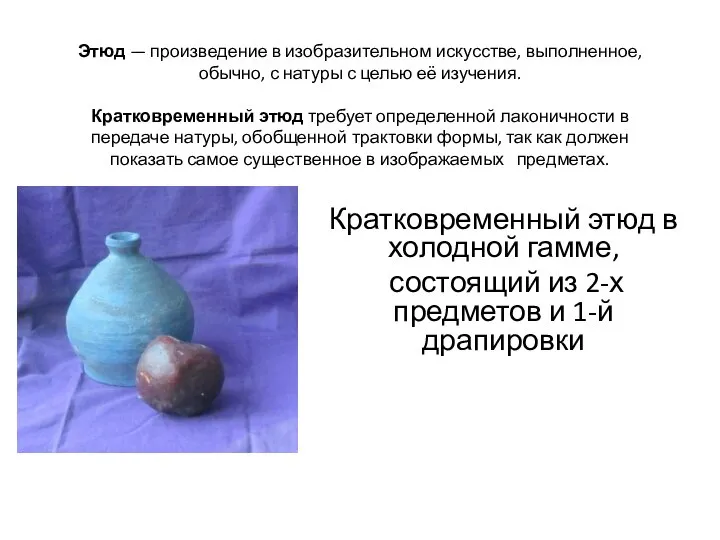 Кратковременный этюд в холодной гамме
 ПРОБЛЕМА КОРРУПЦИЯ В СОВРЕМЕННОЙ РОССИИ
 ПРОБЛЕМА ДЕЛОКАЛИЗАЦИИИСОХРАНЕНИЯ ЗНАНИЯ
 Практическая работа - тест по теме: "Симметрия"
 Презентация на тему Русалочка
  Вторая встреча. Миллион с Аязом 12-13 января
 Ubuntu + Python + Selenium=Легкий Старт
 Автоматизация звука Р в словах и предложениях
  Теоретическое планирование макроциклов, мезоциклов и микроциклов
 Подъёмы, спуск, торможения на лыжах
 Задачи на ноябрь, Почта России
 Презентация на тему Мутации (11 класс)
 20141109_1a._uznay_gornuyu_porodu
 Декодер Delta DMX 4-512
 Презентация на тему Цыгане - нация мира
 A Day to Remember. The Past Simple Tense
 Команда Борцы Экологического Движения
 День защиты детей
 Пункт поиска воевавших родственников
 Инструктаж
 Информационная безопасность: основные понятия и определения