- MeDiMa 3D, digital X-ray tomosynthesis mammography system

Содержание
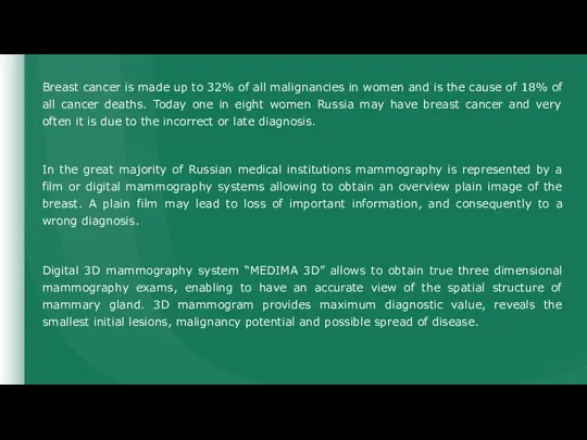
- 2. Breast cancer is made up to 32% of all malignancies in women and is the cause
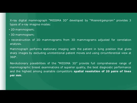
- 3. X-ray digital mammograph “MEDIMA 3D” developed by “Mosrentgenprom” provides 3 types of x-ray imagine modes: 2D
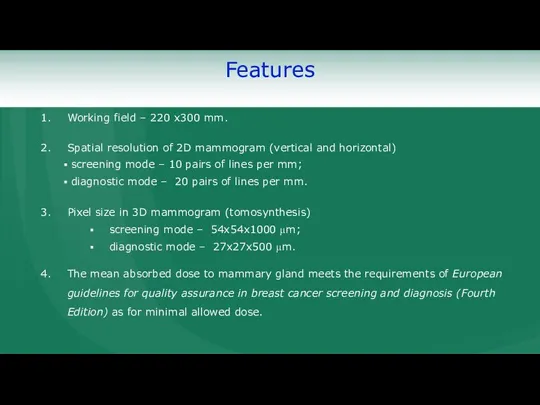
- 4. Features
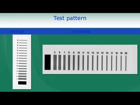
- 5. Test pattern
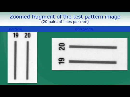
- 6. Zoomed fragment of the test pattern image (20 pairs of lines per mm)
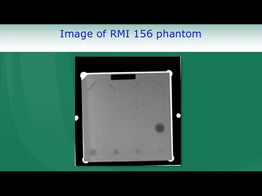
- 9. Image of RMI 156 phantom
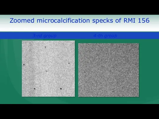
- 10. Zoomed microcalcification specks of RMI 156
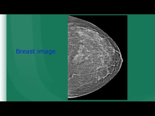
- 11. Breast image
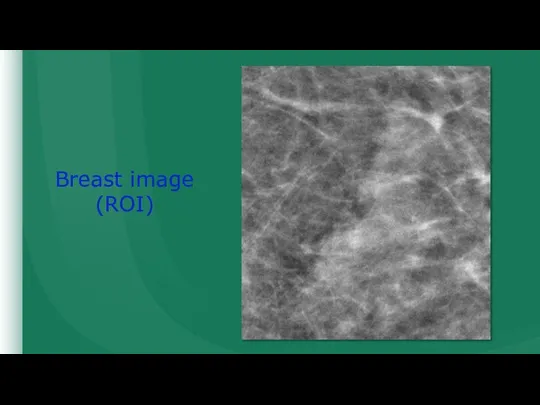
- 12. Breast image (ROI)
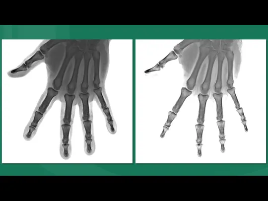
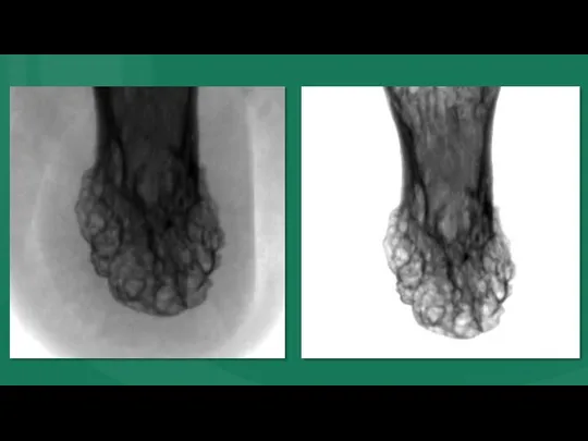
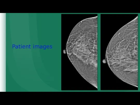
- 13. Patient images
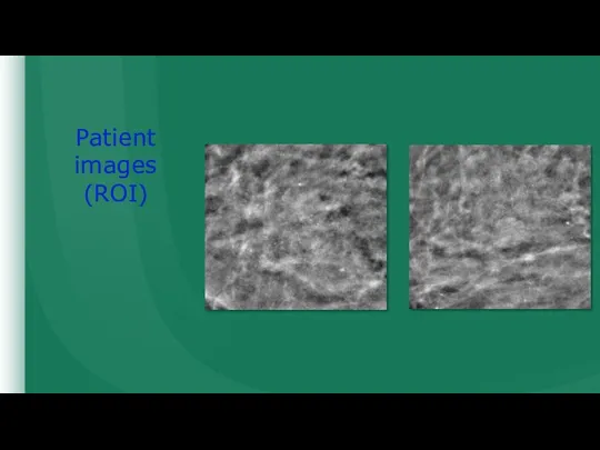
- 14. Patient images (ROI)
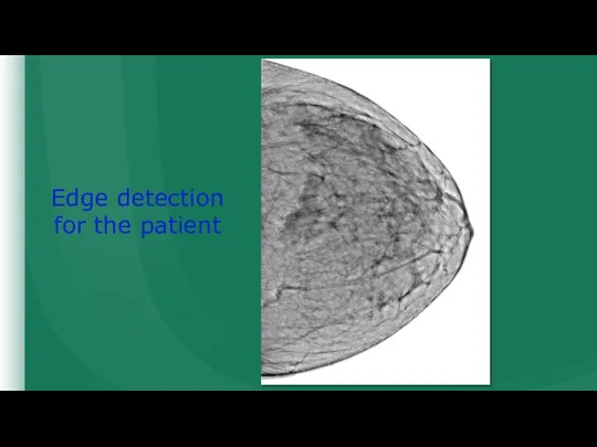
- 15. Edge detection for the patient
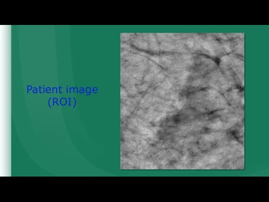
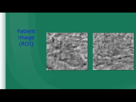
- 16. Patient image (ROI)
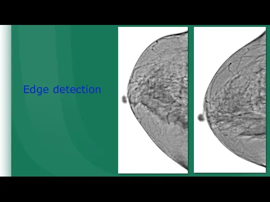
- 17. Edge detection
- 18. Patient image (ROI)
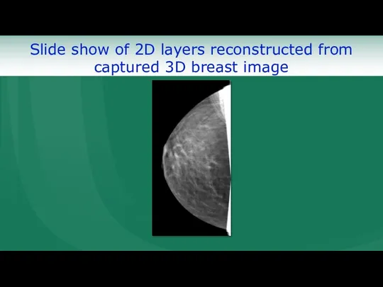
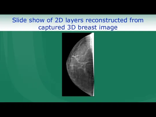
- 19. Slide show of 2D layers reconstructed from captured 3D breast image
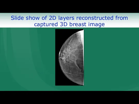
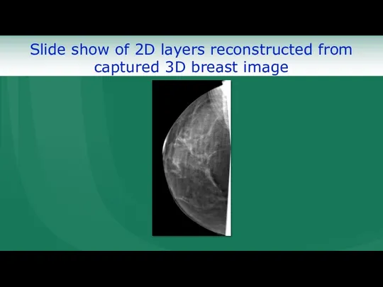
- 20. Slide show of 2D layers reconstructed from captured 3D breast image
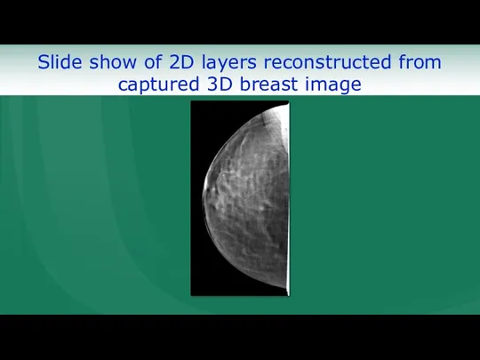
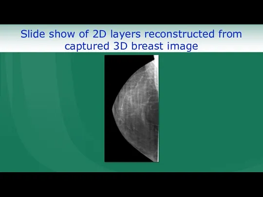
- 21. Slide show of 2D layers reconstructed from captured 3D breast image
- 22. Slide show of 2D layers reconstructed from captured 3D breast image
- 23. Slide show of 2D layers reconstructed from captured 3D breast image
- 24. Slide show of 2D layers reconstructed from captured 3D breast image
- 26. Скачать презентацию
Слайд 3X-ray digital mammograph “MEDIMA 3D” developed by “Mosrentgenprom” provides 3 types of
X-ray digital mammograph “MEDIMA 3D” developed by “Mosrentgenprom” provides 3 types of
Слайд 4Features
Features
Слайд 5Test pattern
Test pattern
Слайд 6Zoomed fragment of the test pattern image
(20 pairs of lines per
Zoomed fragment of the test pattern image
(20 pairs of lines per
Слайд 9Image of RMI 156 phantom
Image of RMI 156 phantom
Слайд 10Zoomed microcalcification specks of RMI 156
Zoomed microcalcification specks of RMI 156
Слайд 11Breast image
Breast image
Слайд 12Breast image
(ROI)
Breast image
(ROI)
Слайд 13Patient images
Patient images
Слайд 14Patient images (ROI)
Patient images (ROI)
Слайд 15Edge detection for the patient
Edge detection for the patient
Слайд 16Patient image (ROI)
Patient image (ROI)
Слайд 17Edge detection
Edge detection
Слайд 18Patient image
(ROI)
Patient image
(ROI)
Слайд 19Slide show of 2D layers reconstructed from captured 3D breast image
Slide show of 2D layers reconstructed from captured 3D breast image
Слайд 20Slide show of 2D layers reconstructed from captured 3D breast image
Slide show of 2D layers reconstructed from captured 3D breast image
Слайд 21Slide show of 2D layers reconstructed from captured 3D breast image
Slide show of 2D layers reconstructed from captured 3D breast image
Слайд 22Slide show of 2D layers reconstructed from captured 3D breast image
Slide show of 2D layers reconstructed from captured 3D breast image
Слайд 23Slide show of 2D layers reconstructed from captured 3D breast image
Slide show of 2D layers reconstructed from captured 3D breast image
Слайд 24Slide show of 2D layers reconstructed from captured 3D breast image
Slide show of 2D layers reconstructed from captured 3D breast image
 Что известно о мозге геймера
 Что надо знать о сезонном гриппе?
 Тема 2. Диспепсический синдром
 Анатомия стопы
 Из истории фагоцитоза
 Клинические формы воспаления
 Адаптивный иммунитет
 Symptoms in cardiovascular diseases
 Наследственные формы КРР
 Введение в специальность. Цели и задачи курса пропедевтики терапевтической стоматологии. Исторические этапы развития
 Осман Илхам
 Болезни геномного импринтинга
 Контролируемый нагрев лазерным излучением внутрикистозной жидкости
 Комплекс упражнений №1. Выполнение движений по тексту
 Сестринский процесс при ревматической лихорадке, пороках сердца. Тема 4.1
 Гигиена детей и подростков
 Отосклероз
 Аменорея, обусловленная нарушением функции коры и гипоталамуса
 Общие вопросы эпидемиологии
 Основные синдромы при патологии желудочно-кишечного тракта
 О качестве оказания медицинской помощи населению
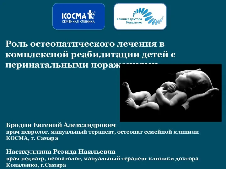 Роль остеопатического лечения в комплексной реабилитации детей с перинатальными поражениями
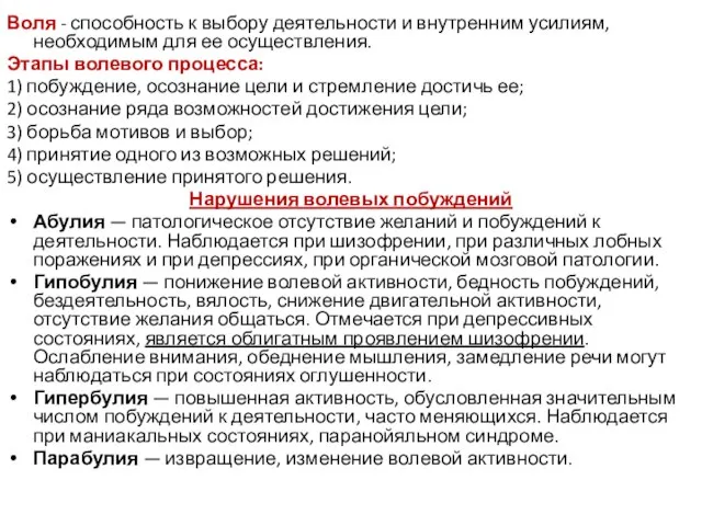 Общая психопатология, лекция6
 Гигиена. 4 класс
 Лекарственные препараты железа (III)
 СПРУ - тяжелое хроническое заболевание
 Первая медицинская помощь при острой сердечной недостаточности и инсульте
 Синдром гипоплазии левых отделов сердца