- Radiological research methods and radiological semiotics of chronic nonspecific lung diseases

Содержание
- 2. Content Radiological methods of chest organs research Radiological anatomy of chest organs Radiological semiotics of chronic
- 3. Radiological research methods chest organs Fluorography Radioscopy Radiography Tomography Bronchography Angiopulmonography Ultrasound diagnostics Computed tomography Magnetic
- 4. The X-ray method is a method of studying the structure and function of various organs and
- 5. Fluorography the method of X-ray examination, which consists in photographing an image from a fluorescent X-ray
- 6. Radioscopy Radioscopy (Greek. scopeo-to consider, observe) is an X-ray examination in which a mobile X-ray image
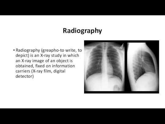
- 7. Radiography Radiography (greapho-to write, to depict) is an X-ray study in which an X-ray image of
- 8. Radiography Overview radiography is an image of the entire organ under study or the entire anatomical
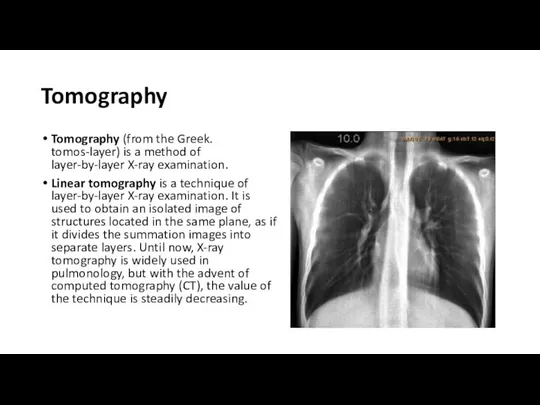
- 9. Tomography Tomography (from the Greek. tomos-layer) is a method of layer-by-layer X-ray examination. Linear tomography is
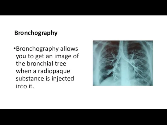
- 10. Bronchography Bronchography allows you to get an image of the bronchial tree when a radiopaque substance
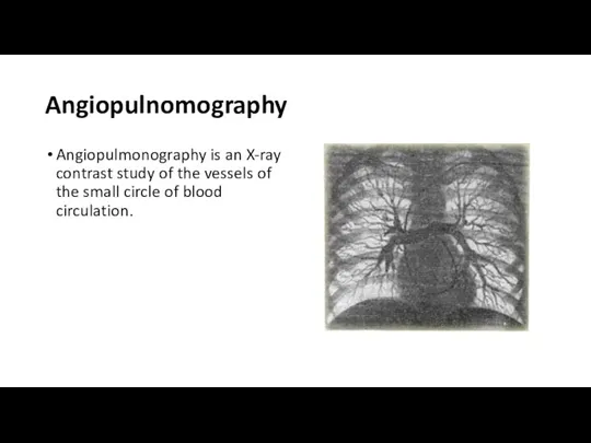
- 11. Angiopulnomography Angiopulmonography is an X-ray contrast study of the vessels of the small circle of blood
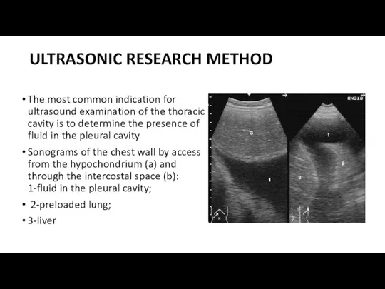
- 12. ULTRASONIC RESEARCH METHOD The most common indication for ultrasound examination of the thoracic cavity is to
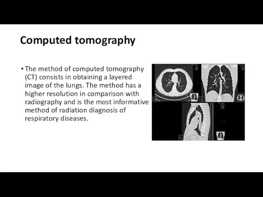
- 13. Computed tomography The method of computed tomography (CT) consists in obtaining a layered image of the
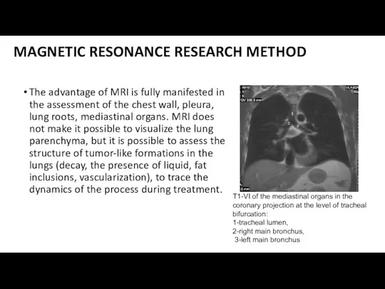
- 14. MAGNETIC RESONANCE RESEARCH METHOD The advantage of MRI is fully manifested in the assessment of the
- 15. RADIONUCLIDE RESEARCH METHOD Radionuclide research methods consist in the introduction into the body (intravenously or by
- 16. Radionuclide methods of lung research are carried out mainly in two versions: - perfusion scintigraphy to
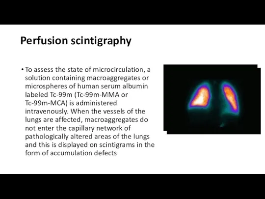
- 17. Perfusion scintigraphy To assess the state of microcirculation, a solution containing macroaggregates or microspheres of human
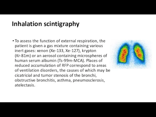
- 18. Inhalation scintigraphy To assess the function of external respiration, the patient is given a gas mixture
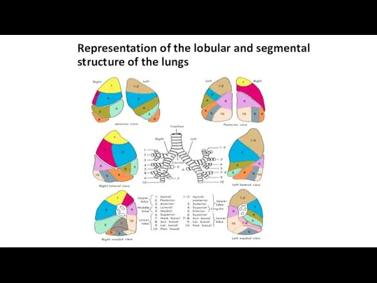
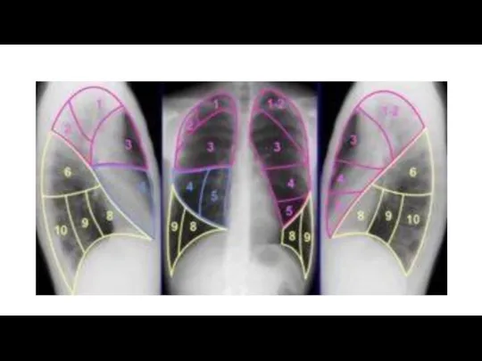
- 19. Representation of the lobular and segmental structure of the lungs
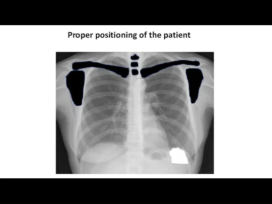
- 20. Proper positioning of the patient
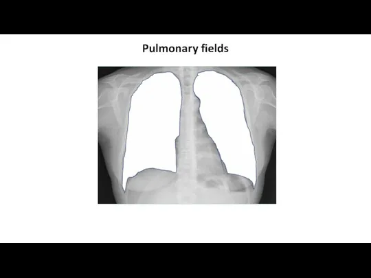
- 21. Pulmonary fields
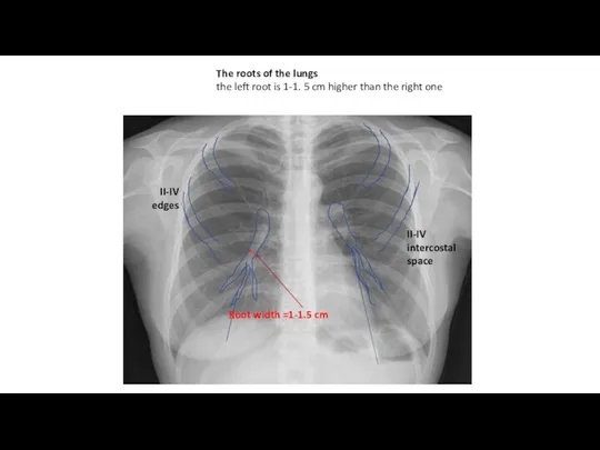
- 22. The roots of the lungs the left root is 1-1. 5 cm higher than the right
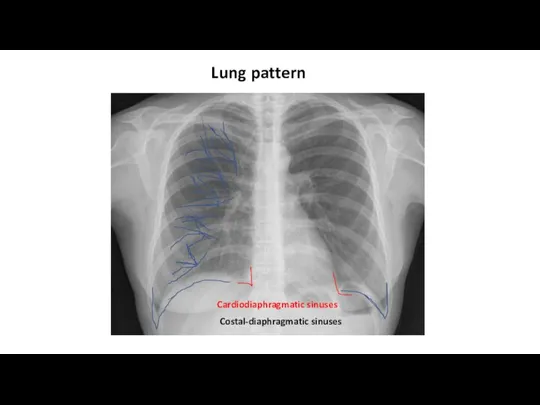
- 23. Costal-diaphragmatic sinuses Lung pattern Cardiodiaphragmatic sinuses
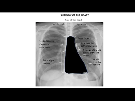
- 24. SHADOW OF THE HEART 1 - Aortic arch / superior vena cava 2-Arc right atrium on
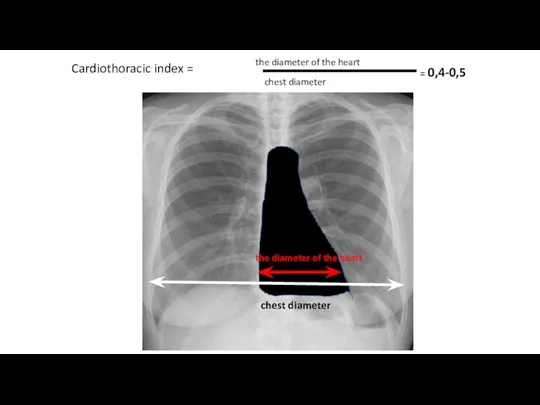
- 25. Cardiothoracic index = the diameter of the heart chest diameter = 0,4-0,5 the diameter of the
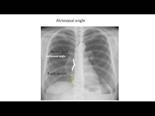
- 26. Atriovasal angle Aortic arch Right atrium Atriovasal angle
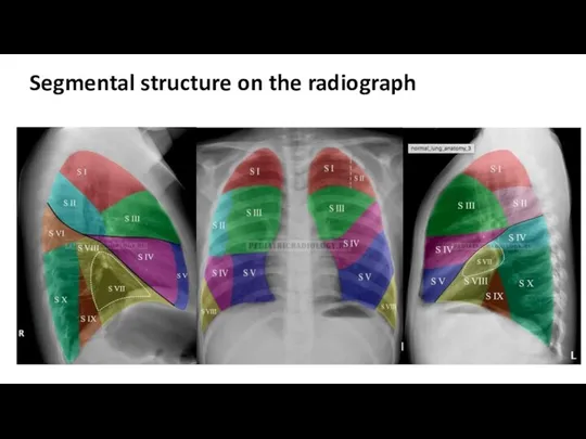
- 27. Segmental structure on the radiograph
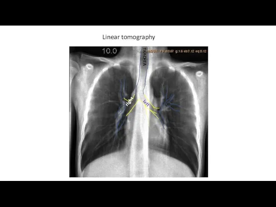
- 29. Linear tomography TRACHEA right left
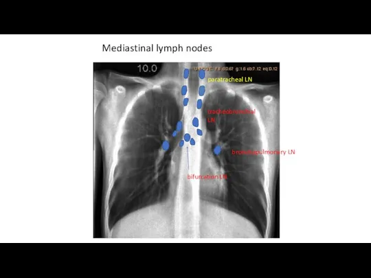
- 30. Mediastinal lymph nodes paratracheal LN bronchopulmonary LN tracheobronchial LN bifurcation LN
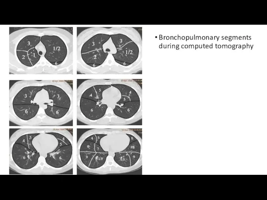
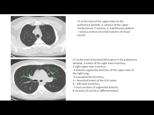
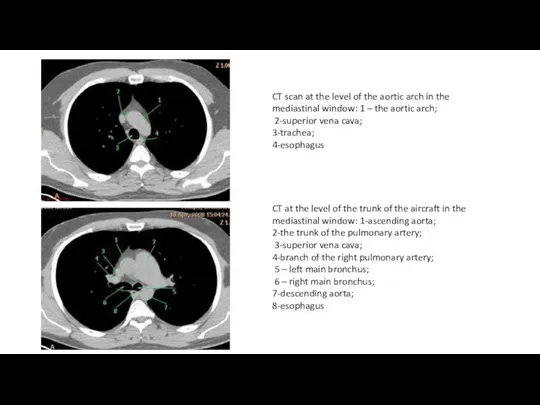
- 31. Bronchopulmonary segments during computed tomography
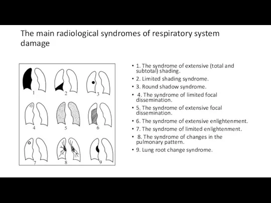
- 34. The main radiological syndromes of respiratory system damage 1. The syndrome of extensive (total and subtotal)
- 35. Chronic nonspecific lung diseases Chronic nonspecific lung diseases(CNL), a group of chronic diseases of the bronchopulmonary
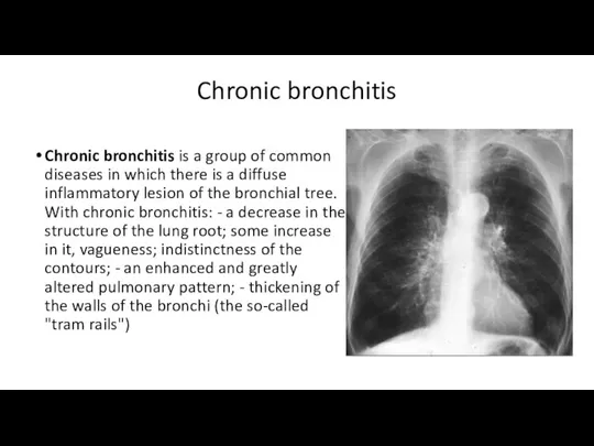
- 36. Chronic bronchitis Chronic bronchitis is a group of common diseases in which there is a diffuse
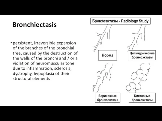
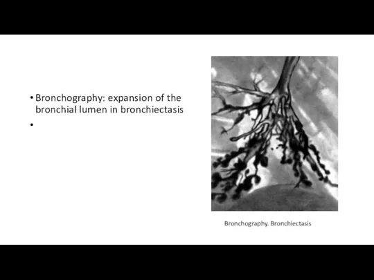
- 37. Bronchiectasis persistent, irreversible expansion of the branches of the bronchial tree, caused by the destruction of
- 38. Bronchography: expansion of the bronchial lumen in bronchiectasis Bronchography. Bronchiectasis
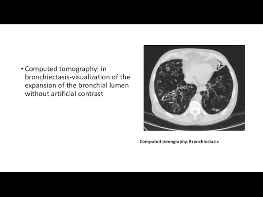
- 39. Computed tomography: in bronchiectasis-visualization of the expansion of the bronchial lumen without artificial contrast Computed tomography.
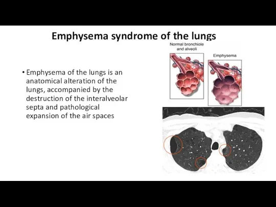
- 40. Emphysema syndrome of the lungs Emphysema of the lungs is an anatomical alteration of the lungs,
- 41. According to the degree of involvement in the pathological process of acinus, the following types of
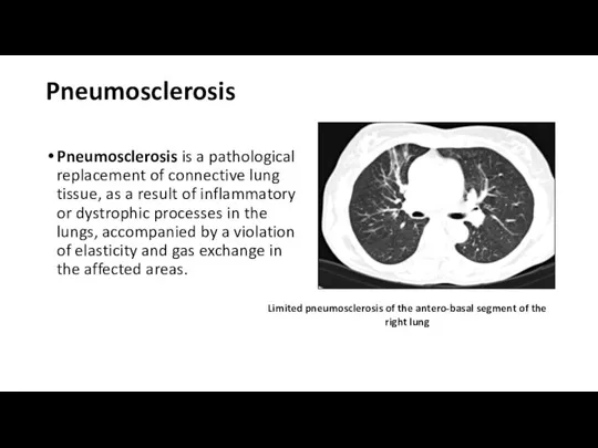
- 42. Pneumosclerosis Pneumosclerosis is a pathological replacement of connective lung tissue, as a result of inflammatory or
- 43. According to the degree of severity of the replacement of the pulmonary parenchyma with connective tissue,
- 44. CHRONIC NONSPECIFIC PNEUMONIA a limited inflammatory process of the lungs, characterized by the development of purulent-necrotic
- 46. Скачать презентацию
Слайд 2Content
Radiological methods of chest organs
research
Radiological anatomy of chest organs
Radiological
Content
Radiological methods of chest organs
research
Radiological anatomy of chest organs
Radiological
Слайд 3Radiological research methods
chest organs
Fluorography
Radioscopy
Radiography
Tomography
Bronchography
Angiopulmonography
Ultrasound diagnostics
Computed
Radiological research methods
chest organs
Fluorography
Radioscopy
Radiography
Tomography
Bronchography
Angiopulmonography
Ultrasound diagnostics
Computed
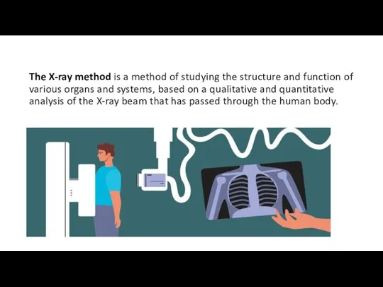
Слайд 4The X-ray method is a method of studying the structure and function
The X-ray method is a method of studying the structure and function
Слайд 5Fluorography
the method of X-ray examination, which consists in photographing an image from
Fluorography
the method of X-ray examination, which consists in photographing an image from
Слайд 6Radioscopy
Radioscopy (Greek. scopeo-to consider, observe) is an X-ray examination in which a
Radioscopy
Radioscopy (Greek. scopeo-to consider, observe) is an X-ray examination in which a
Слайд 7Radiography
Radiography (greapho-to write, to depict) is an X-ray study in which an
Radiography
Radiography (greapho-to write, to depict) is an X-ray study in which an
Слайд 8Radiography
Overview radiography is an image of the entire organ under study or
Radiography
Overview radiography is an image of the entire organ under study or
Слайд 9Tomography
Tomography (from the Greek. tomos-layer) is a method of layer-by-layer X-ray examination.
Linear
Tomography
Tomography (from the Greek. tomos-layer) is a method of layer-by-layer X-ray examination.
Linear
Слайд 10
Bronchography
Bronchography allows you to get an image of the bronchial tree
Bronchography
Bronchography allows you to get an image of the bronchial tree
Слайд 11Angiopulnomography
Angiopulmonography is an X-ray contrast study of the vessels of the small
Angiopulnomography
Angiopulmonography is an X-ray contrast study of the vessels of the small
Слайд 12ULTRASONIC RESEARCH METHOD
The most common indication for ultrasound examination of the thoracic
ULTRASONIC RESEARCH METHOD
The most common indication for ultrasound examination of the thoracic
Слайд 13Computed tomography
The method of computed tomography (CT) consists in obtaining a layered
Computed tomography
The method of computed tomography (CT) consists in obtaining a layered
Слайд 14MAGNETIC RESONANCE RESEARCH METHOD
The advantage of MRI is fully manifested in the
MAGNETIC RESONANCE RESEARCH METHOD
The advantage of MRI is fully manifested in the
Слайд 15RADIONUCLIDE RESEARCH METHOD
Radionuclide research methods consist in the introduction into the body
RADIONUCLIDE RESEARCH METHOD
Radionuclide research methods consist in the introduction into the body
Слайд 16Radionuclide methods of lung research are carried out mainly in two versions:
-
Radionuclide methods of lung research are carried out mainly in two versions:
-
Слайд 17Perfusion scintigraphy
To assess the state of microcirculation, a solution containing macroaggregates or
Perfusion scintigraphy
To assess the state of microcirculation, a solution containing macroaggregates or
Слайд 18Inhalation scintigraphy
To assess the function of external respiration, the patient is given
Inhalation scintigraphy
To assess the function of external respiration, the patient is given
Слайд 19Representation of the lobular and segmental structure of the lungs
Representation of the lobular and segmental structure of the lungs
Слайд 20Proper positioning of the patient
Proper positioning of the patient
Слайд 21Pulmonary fields
Pulmonary fields
Слайд 22The roots of the lungs
the left root is 1-1. 5 cm higher
The roots of the lungs the left root is 1-1. 5 cm higher
Слайд 23Costal-diaphragmatic sinuses
Lung pattern
Cardiodiaphragmatic sinuses
Costal-diaphragmatic sinuses
Lung pattern
Cardiodiaphragmatic sinuses
Слайд 24SHADOW OF THE HEART
1 - Aortic arch / superior vena cava
2-Arc right
SHADOW OF THE HEART
1 - Aortic arch / superior vena cava
2-Arc right
Слайд 25Cardiothoracic index =
the diameter of the heart
chest diameter
= 0,4-0,5
the diameter of the
Cardiothoracic index =
the diameter of the heart
chest diameter
= 0,4-0,5
the diameter of the
Слайд 26Atriovasal angle
Aortic arch
Right atrium
Atriovasal angle
Atriovasal angle
Aortic arch
Right atrium
Atriovasal angle
Слайд 27Segmental structure on the radiograph
Segmental structure on the radiograph
Слайд 29Linear tomography
TRACHEA
right
left
Linear tomography
TRACHEA
right
left
Слайд 30Mediastinal lymph nodes
paratracheal LN
bronchopulmonary LN
tracheobronchial LN
bifurcation LN
Mediastinal lymph nodes
paratracheal LN
bronchopulmonary LN
tracheobronchial LN
bifurcation LN
Слайд 31Bronchopulmonary segments during computed tomography
Bronchopulmonary segments during computed tomography
Слайд 34The main radiological syndromes of respiratory system damage
1. The syndrome of extensive
The main radiological syndromes of respiratory system damage
1. The syndrome of extensive
Слайд 35Chronic nonspecific lung diseases
Chronic nonspecific lung diseases(CNL), a group of chronic diseases
Chronic nonspecific lung diseases
Chronic nonspecific lung diseases(CNL), a group of chronic diseases
Слайд 36Chronic bronchitis
Chronic bronchitis is a group of common diseases in which there
Chronic bronchitis
Chronic bronchitis is a group of common diseases in which there
Слайд 37Bronchiectasis
persistent, irreversible expansion of the branches of the bronchial tree, caused by
Bronchiectasis
persistent, irreversible expansion of the branches of the bronchial tree, caused by
Слайд 38Bronchography: expansion of the bronchial lumen in bronchiectasis
Bronchography. Bronchiectasis
Bronchography: expansion of the bronchial lumen in bronchiectasis
Bronchography. Bronchiectasis
Слайд 39Computed tomography: in bronchiectasis-visualization of the expansion of the bronchial lumen without
Computed tomography: in bronchiectasis-visualization of the expansion of the bronchial lumen without
Слайд 40Emphysema syndrome of the lungs
Emphysema of the lungs is an anatomical alteration
Emphysema syndrome of the lungs
Emphysema of the lungs is an anatomical alteration
Слайд 41According to the degree of involvement in the pathological process of acinus,
According to the degree of involvement in the pathological process of acinus,
Слайд 42Pneumosclerosis
Pneumosclerosis is a pathological replacement of connective lung tissue, as a result
Pneumosclerosis
Pneumosclerosis is a pathological replacement of connective lung tissue, as a result
Слайд 43According to the degree of severity of the replacement of the pulmonary
According to the degree of severity of the replacement of the pulmonary
Слайд 44CHRONIC NONSPECIFIC PNEUMONIA
a limited inflammatory process of the lungs, characterized by the
CHRONIC NONSPECIFIC PNEUMONIA
a limited inflammatory process of the lungs, characterized by the
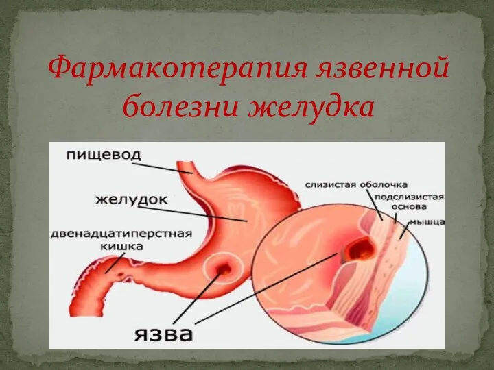 Фармакотерапия язвенной болезни желудка
 Предмет и задачи возрастной физиологии. (Лекция 1)
 Методика приготовления нативных и окрашенных препаратов мокроты
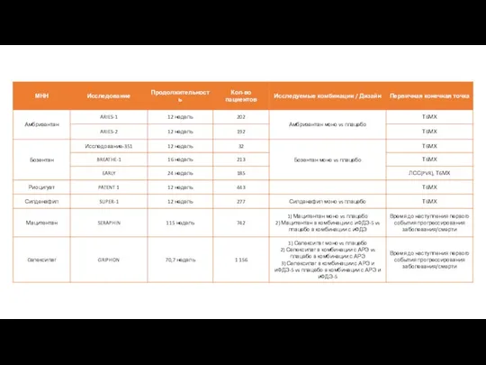 Описание исследований лекарственных препаратов
 Emergencies in children endocrinology
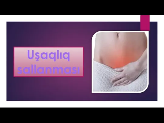 Uşaqlıq sallanması
 Современные принципы лечения аутоиммунной патологии
 Местная анестезия
 Бүйрек және эндокринді жүйе ауруларындағы неврологиялық синдромдар
 Расспрос, осмотр, пальпация сердечной области. Лекция 6
 Резекционные операции на теле и хвосте поджелудочной железы, тотальная панкреатэктомия
 Синоатриальные и атриовентрикулярные блокады: причины, экг-диагностика, симптоматика
 Психовегетативные жалобы в практике участкового врача
 Чесотка в аспекте инфекций, передаваемых половым путем (по материалам Смоленской области)
 Диагностика, профилактика и оздоровительные мероприятия при ЭМКАРе и брадзоте рогатого скота
 Исследование ассортимента и потребительских предпочтений лекарственных препаратов на основе амброксола
 Гипоксия. Гипоксические состояния
 Питание как компонент терапии респираторных инфекций у детей, включая COVID-19. Вопросы лечебного питания при ОКИ
 Аутизм, РАС-биокоррекция при аутизме и РАС. Интенсив для родителей. Занятие 3
 Пародонтит
 Внесення інформації в ЕСОЗ ЩОДО вакцинації від covid-19
 Когнитивно - поведенческое направление в психотерапии. Рациональная психотерапия Дюбуа
 Столбняк. Признаки столбняка
 Практикум по ЭКГ
 Лаборатория Большая Перемена
 Центр эстетического взаимодействия студентов МарГУ
 Профилактика туберкулеза
 Понятие перелома. Клиника. Диагностика на догоспитальном этапе