- Hypertrophic cardiomyopathy

Содержание
- 2. Plan: Classification Definition of disease Etiology Morphology: - macro image; - micro image. Complications Conclusion Reference
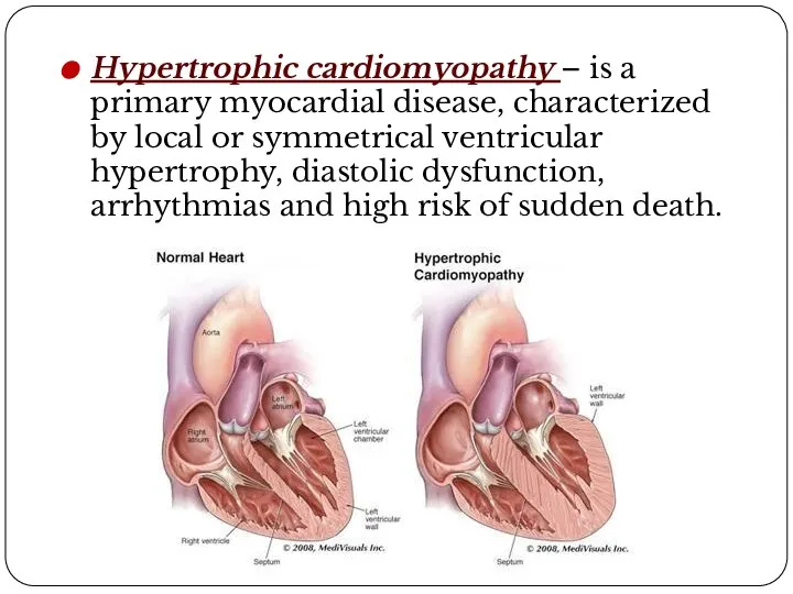
- 4. Hypertrophic cardiomyopathy – is a primary myocardial disease, characterized by local or symmetrical ventricular hypertrophy, diastolic
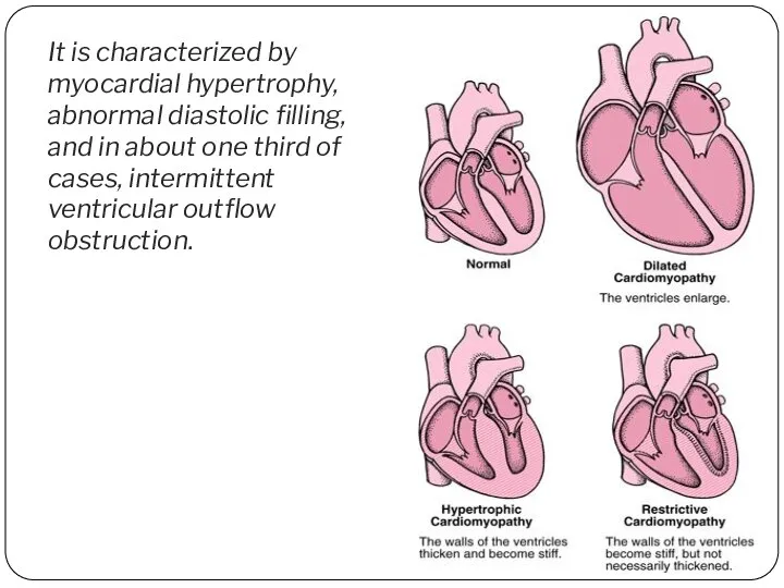
- 5. It is characterized by myocardial hypertrophy, abnormal diastolic filling, and in about one third of cases,
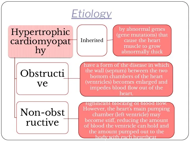
- 6. Etiology have a form of the disease in which the wall (septum) between the two bottom
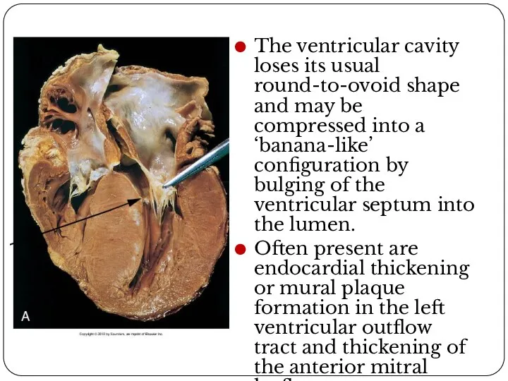
- 7. Macro image The ventricular cavity loses its usual round-to-ovoid shape and may be compressed into a
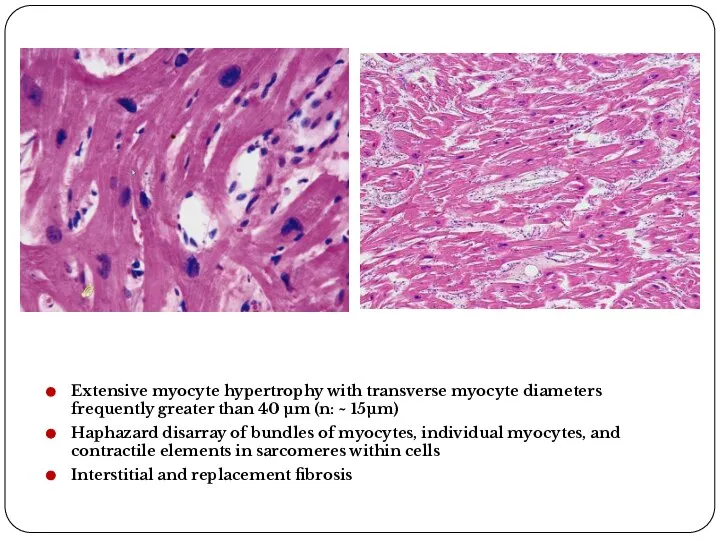
- 8. Extensive myocyte hypertrophy with transverse myocyte diameters frequently greater than 40 μm (n: ~ 15μm) Haphazard
- 9. Literature: V.Kumar, A.K. Abbas, S.N. Fauso. Pathologic Basis of Disease, 7th edition, 2008 – 1525 p.
- 11. Скачать презентацию
Слайд 2Plan:
Classification
Definition of disease
Etiology
Morphology:
- macro image;
- micro image.
Complications
Conclusion
Reference
Plan:
Classification
Definition of disease
Etiology
Morphology:
- macro image;
- micro image.
Complications
Conclusion
Reference
Слайд 4Hypertrophic cardiomyopathy – is a primary myocardial disease, characterized by local or
Hypertrophic cardiomyopathy – is a primary myocardial disease, characterized by local or
Слайд 5It is characterized by myocardial hypertrophy, abnormal diastolic filling, and in about
It is characterized by myocardial hypertrophy, abnormal diastolic filling, and in about
Слайд 6Etiology
have a form of the disease in which the wall (septum) between
Etiology
have a form of the disease in which the wall (septum) between
Слайд 7Macro image
The ventricular cavity loses its usual round-to-ovoid shape and may be
Macro image
The ventricular cavity loses its usual round-to-ovoid shape and may be
Слайд 8Extensive myocyte hypertrophy with transverse myocyte diameters frequently greater than 40 μm
Extensive myocyte hypertrophy with transverse myocyte diameters frequently greater than 40 μm
Слайд 9Literature:
V.Kumar, A.K. Abbas, S.N. Fauso. Pathologic Basis of Disease, 7th edition, 2008
Literature:
V.Kumar, A.K. Abbas, S.N. Fauso. Pathologic Basis of Disease, 7th edition, 2008
 Хроническая почечная недост
 Гендік инженерия
 Актуальные вопросы профилактики, диагностики и лечения коронавирусной инфекции Сovid-19
 Новоутворення яєчників після гістеректомії: діагностика і тактика оперативного лікування
 Кинезотерапия. Виды кинезотерапии. Показания для применения
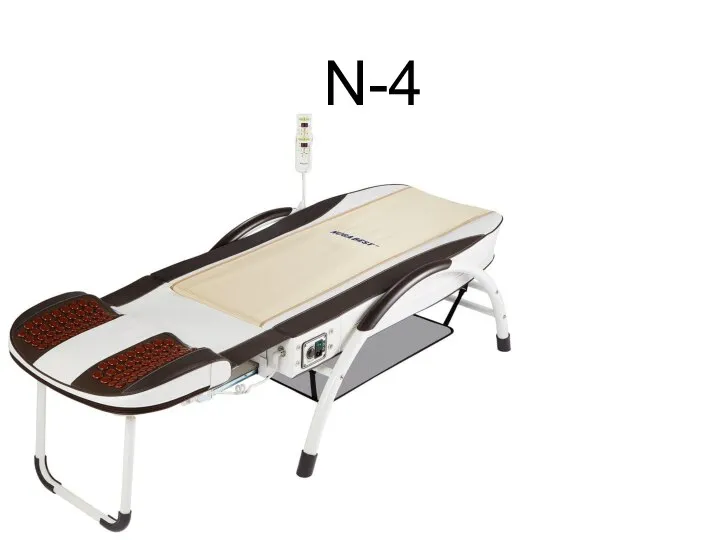 Массажное оборудование N-4
 Получение спермы от быков производителей. Хранение, транспортировка спермы
 Первая помощь при передозировке психотропных веществ
 Синдром Прадера-Вилли
 Микроцефалия
 Гнойные воспаления костей
 ҚР ДСМ ТКҚСҚБК ҰСО ШЖҚ РМК Түркістан облысы вирусологиялық зертханасы
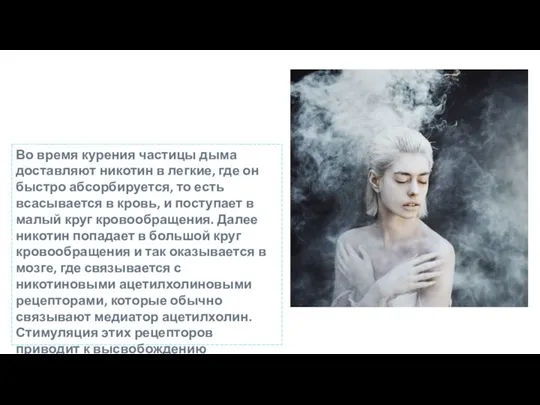 Курение
 Лучевая семиотика при заболеваниях зубочелюстной системы. 2 часть
 Здоровое питание. Принципы здорового питания: это должен знать каждый!
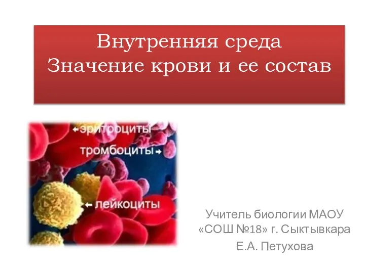 Внутренняя среда. Значение крови и ее состав
 Медицинская микробиология
 Мозг и сознание
 ძუძუს დაავადებები
 Клиника ДВС синдрома
 Травмы сердца
 Оборудование реанимационной палаты
 Научно-практический календарь травматологии и ортопедии
 Кафедра детских болезней педиатрического факультета
 Индивидуальный подбор при расстройствах регуляции РС. Лекция 16
 Служба профилактики повторных переломов в РФ
 Гипсование композиции в кювету. Выпаривание воска
 Заболевания конъюктивы