Serum rantes, transforming growth factor-β1 and interleukin-6 fibrosis in patients with aortic valve stenosis
- Serum rantes, transforming growth factor-β1 and interleukin-6 fibrosis in patients with aortic valve stenosis

Содержание
- 2. INTRODUCTION Progressive aortic valve degeneration leads to severe aortic valve stenosis (AS) in approximately 2 –
- 3. Some postulated factors driving AS progession include influence of classic atherosclerotic risk factors. In AS, not
- 4. Myocardial fibrosis results from increased myofibroblast activity and excessive extracellular matrix deposition. Various cells and molecules
- 5. METHODS Study population Magnetic resonance imaging Inflammatory biomarkers Echocardiography Statistical analysis
- 6. Study population Forty consecutive patients with moderate (defined as an aortic valve area between 1.0 –
- 7. Magnetic resonance imaging LV end-diastolic volume and diameters, LV end-systolic volume and diameters, LV ejection fraction
- 8. Inflammatory biomarkers Fasting blood was drawn from an antecubital vein without tourniquet and placed in a
- 9. Echocardiography Comprehensive transthoracic echocardiography was performed in all patients after ≥30 minutes of rest by 2
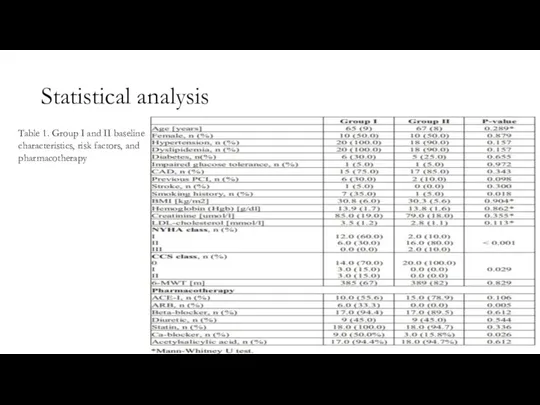
- 10. Statistical analysis Table 1. Group I and II baseline characteristics, risk factors, and pharmacotherapy
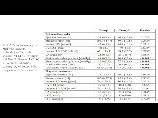
- 11. Table 2. Echocardiography and MRI measurements. Abbreviations: SV, stroke volume; LVEDD, left ventricle end diastolic diameter;
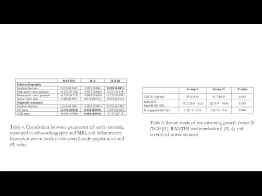
- 12. Table 3. Serum levels of transforming growth factor β1 (TGF-β1), RANTES and interleukin 6 (IL-6) and
- 13. RESULTS Group I included twenty patients with moderate AS while group II included twenty patients with
- 14. In previous studies, significant differences in serum levels of TGF-β1 were found in severe AS patients
- 15. Conclusions Although there is an increasing interest in the immunopathogenesis of AS, relatively little is known
- 17. Скачать презентацию
Слайд 2INTRODUCTION
Progressive aortic valve degeneration leads to severe aortic valve stenosis (AS) in
INTRODUCTION
Progressive aortic valve degeneration leads to severe aortic valve stenosis (AS) in
Слайд 3Some postulated factors driving AS progession include influence of classic atherosclerotic risk
Some postulated factors driving AS progession include influence of classic atherosclerotic risk
Слайд 4Myocardial fibrosis results from increased myofibroblast activity and excessive extracellular matrix deposition.
Myocardial fibrosis results from increased myofibroblast activity and excessive extracellular matrix deposition.
Слайд 5METHODS
Study population
Magnetic resonance imaging
Inflammatory biomarkers
Echocardiography
Statistical analysis
METHODS
Study population
Magnetic resonance imaging
Inflammatory biomarkers
Echocardiography
Statistical analysis
Слайд 6Study population
Forty consecutive patients with moderate (defined as an aortic valve area
Study population
Forty consecutive patients with moderate (defined as an aortic valve area
Слайд 7Magnetic resonance imaging
LV end-diastolic volume and diameters, LV end-systolic volume and diameters,
Magnetic resonance imaging
LV end-diastolic volume and diameters, LV end-systolic volume and diameters,
Слайд 8Inflammatory biomarkers
Fasting blood was drawn from an antecubital vein without tourniquet and
Inflammatory biomarkers
Fasting blood was drawn from an antecubital vein without tourniquet and
Слайд 9Echocardiography
Comprehensive transthoracic echocardiography was performed in all patients after ≥30 minutes of
Echocardiography
Comprehensive transthoracic echocardiography was performed in all patients after ≥30 minutes of
Слайд 10Statistical analysis
Table 1. Group I and II baseline characteristics, risk factors, and
Statistical analysis
Table 1. Group I and II baseline characteristics, risk factors, and
Слайд 11Table 2. Echocardiography and MRI measurements. Abbreviations: SV, stroke volume; LVEDD, left
Table 2. Echocardiography and MRI measurements. Abbreviations: SV, stroke volume; LVEDD, left
Слайд 12Table 3. Serum levels of transforming growth factor β1 (TGF-β1), RANTES and
Table 3. Serum levels of transforming growth factor β1 (TGF-β1), RANTES and
Слайд 13RESULTS
Group I included twenty patients with moderate AS while group II included
RESULTS
Group I included twenty patients with moderate AS while group II included
Слайд 14In previous studies, significant differences in serum levels of TGF-β1 were found
In previous studies, significant differences in serum levels of TGF-β1 were found
Слайд 15Conclusions
Although there is an increasing interest in the immunopathogenesis of AS, relatively
Conclusions
Although there is an increasing interest in the immunopathogenesis of AS, relatively
 pptx лихорадка ласса.pptxahmad
 Mécanismes biochimiques du développement des pathologies des tissus de la cavité buccale
 Роль медицинской сестры дошкольного учреждения в профилактике заболеваний у дошкольников
 Ишемическая болезнь сердца (ИБС). Стенокардия
 Канцерогенные факторы окружающей среды
 Презентация по психологии на тему _Психологический профиль онкобольных_
 Клиническая фармакология ингаляционных анестетиков: Изофлуран и Севофлуран
 Оң қарыншаның миокард инфарктының ЭКГ белгілері
 Как сохранить зрение
 статучет 2022 МИАЦ
 Дифференциальная диагностика опухолей и воспалительных заболеваний легких
 Методы идентификации клеточных элементов в моче: суправитальная окраска препаратов осадка мочи, подсчёт уролейкограммы
 Принципы рациональной антибиотикотерапии у детей
 Презентация по иммунологии на тему Туберкулез
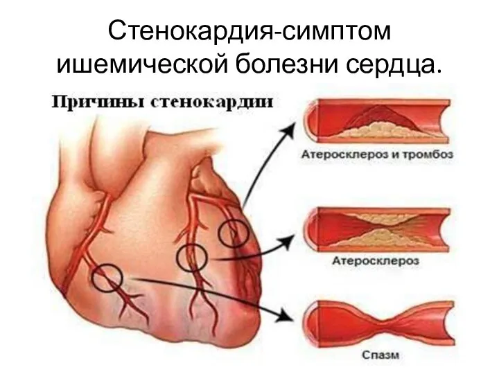 Стенокардия - симптом ишемической болезни сердца
 Инфузионная терапия. Забор крови для лабораторных исследований
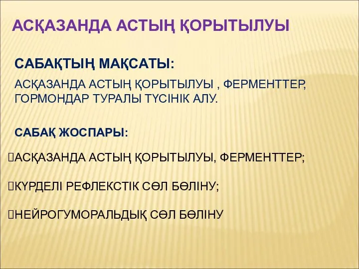 Асқазанда астың қорытылуы, ферменттер, гормондар туралы түсінік алу
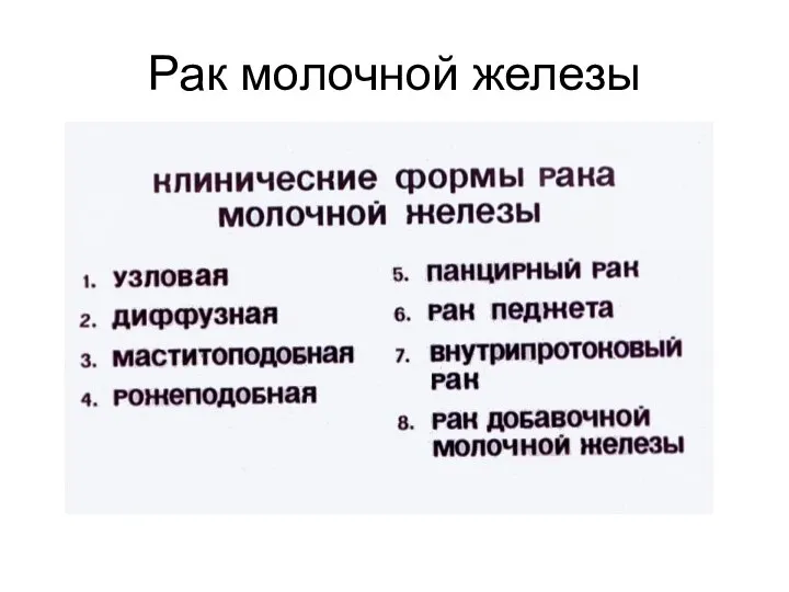 Рак молочной железы
 Знание - ответственность - здоровье. Вирусы
 Некоторые проблемы и решения на примере ВИЧ-инфекции и алкоголя
 Тенденция развития ВИЧ-инфекции в России и Брянской области
 Органные дисфункции
 Дермотологияда ГКС терапиясы
 Профессионализм медицинской сестры. Свойства, функции, признаки
 Принципы оздоровления организма
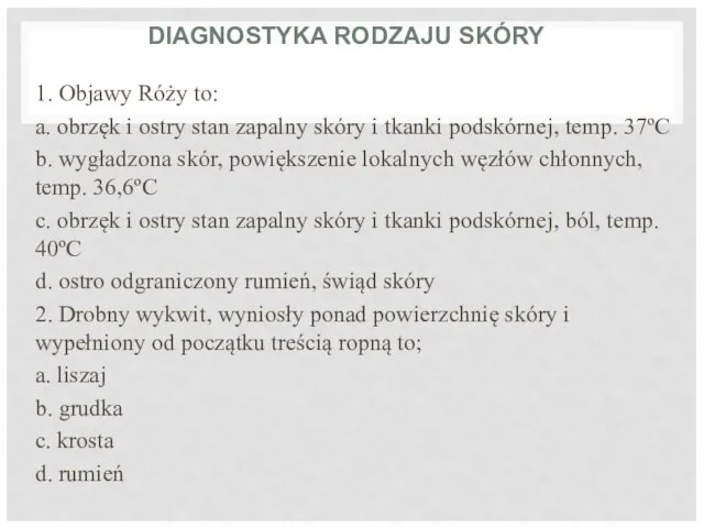 Diagnostyka rodzaju skóry (тест)
 Развитие детей раннего младенческого возраста в онтогенезе
 Аллергия. Реакции III типа – иммунокомплексные