- Common principles of purulent surgery

Содержание
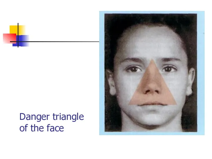
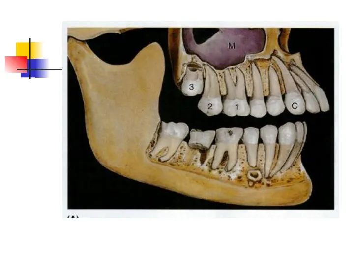
- 5. Danger triangle of the face
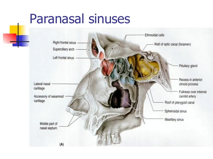
- 7. Paranasal sinuses
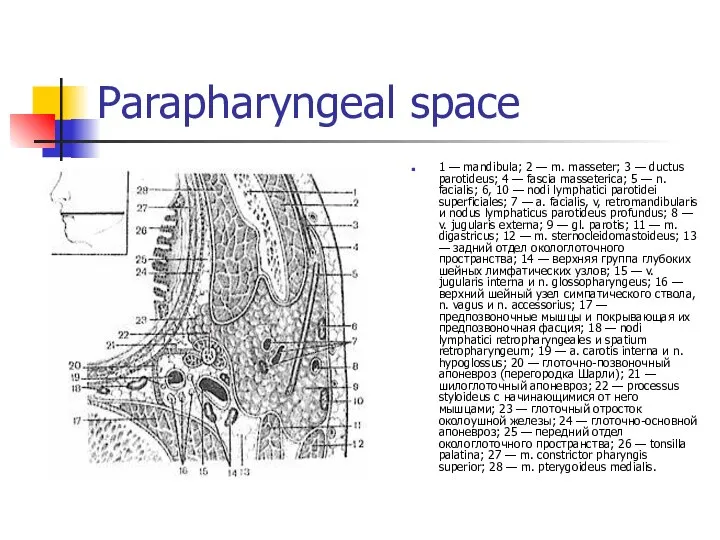
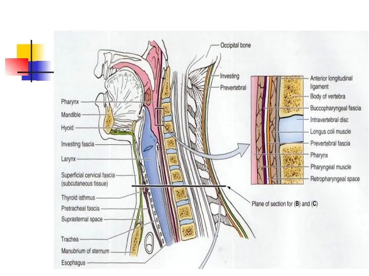
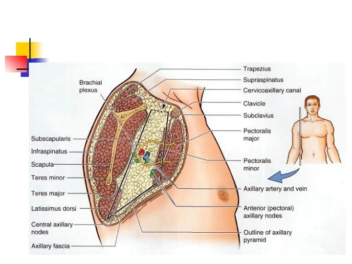
- 8. Parapharyngeal space 1 — mandibula; 2 — m. masseter; 3 — ductus parotideus; 4 — fascia
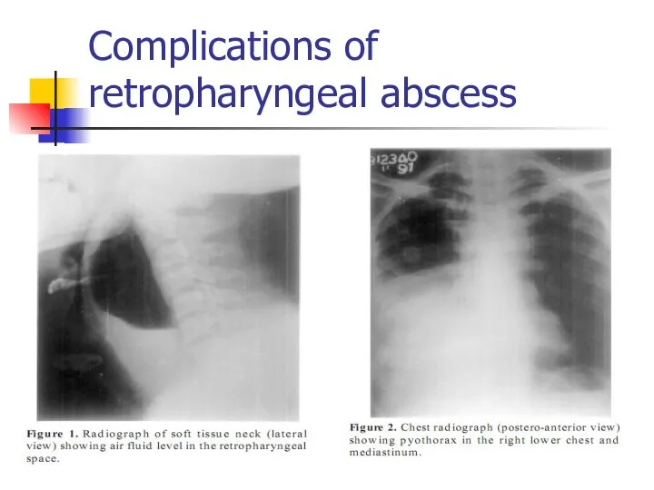
- 11. Complications of retropharyngeal abscess
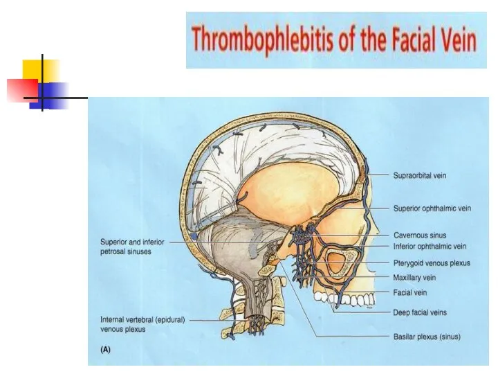
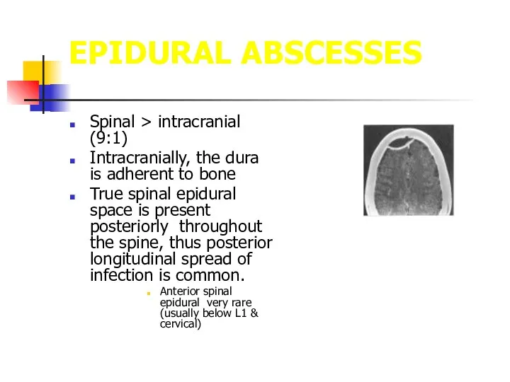
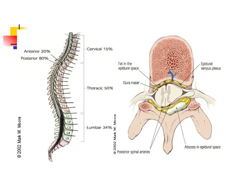
- 12. EPIDURAL ABSCESSES Spinal > intracranial (9:1) Intracranially, the dura is adherent to bone True spinal epidural
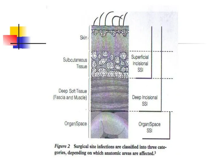
- 13. American Family Physician April 1, 2002
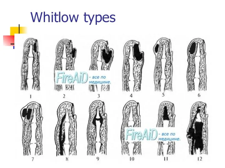
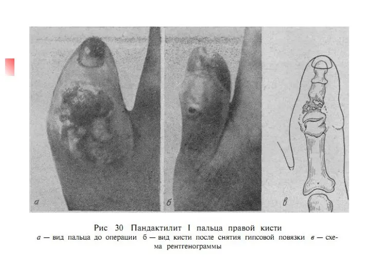
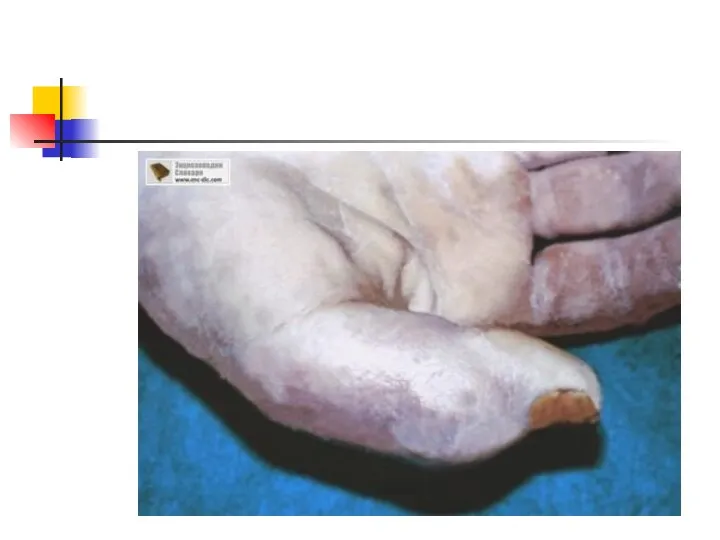
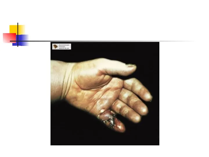
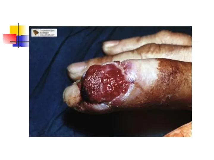
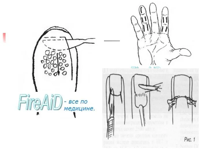
- 16. Whitlow types
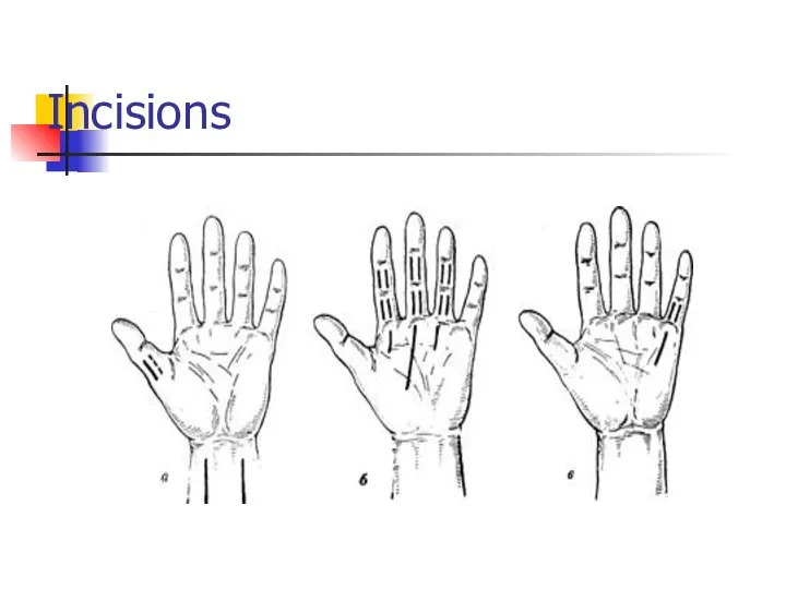
- 27. Incisions
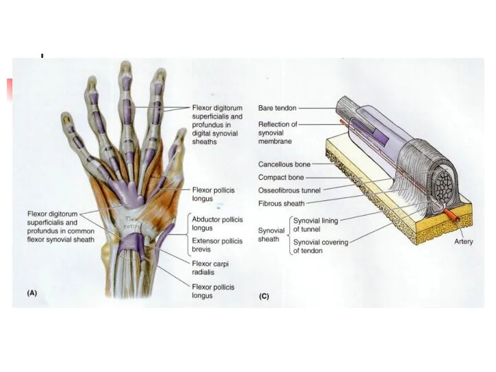
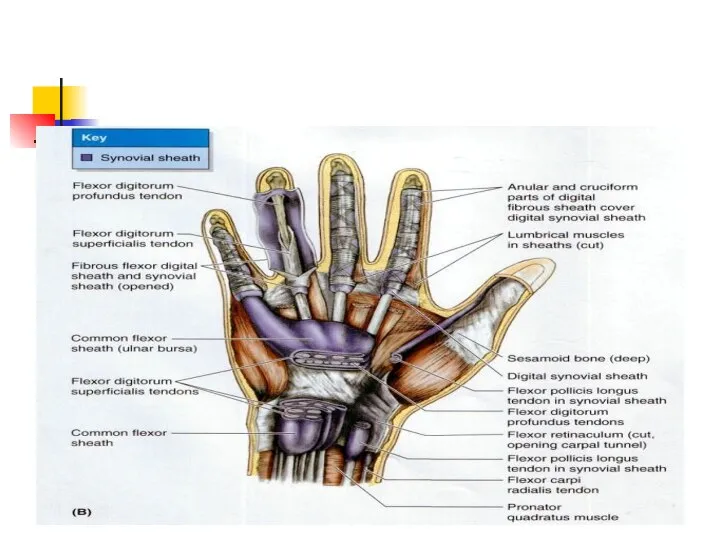
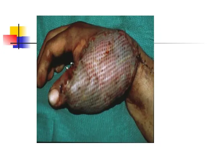
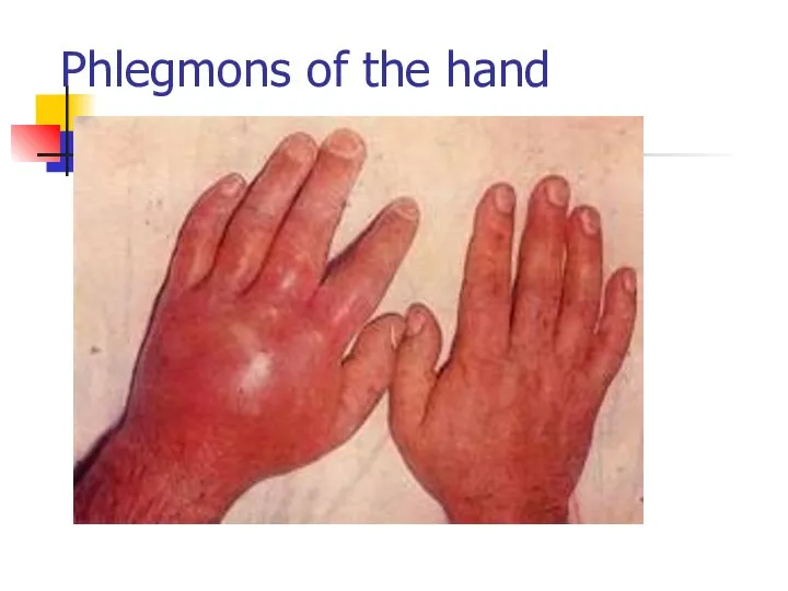
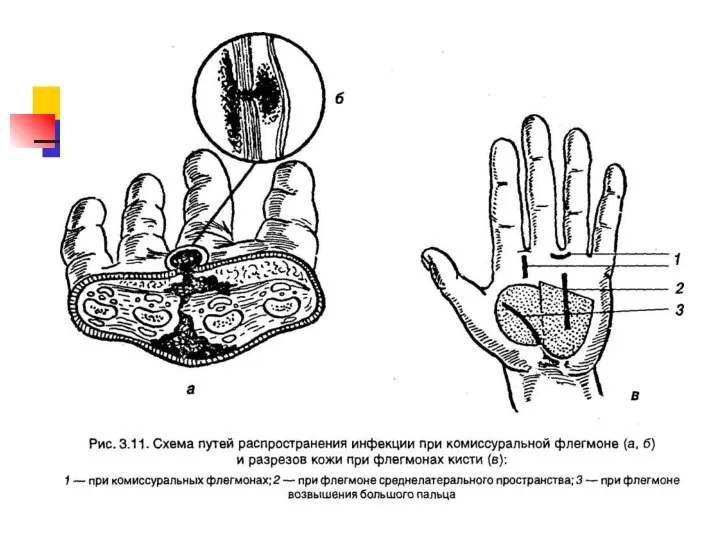
- 28. Phlegmons of the hand
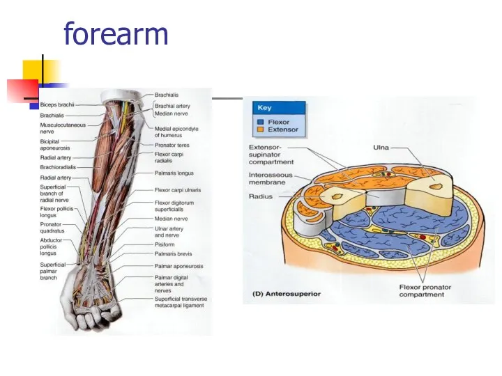
- 30. forearm
- 46. Скачать презентацию
Слайд 5Danger triangle of the face
Danger triangle of the face
Слайд 7Paranasal sinuses
Paranasal sinuses
Слайд 8Parapharyngeal space
1 — mandibula; 2 — m. masseter; 3 — ductus parotideus;
Parapharyngeal space
1 — mandibula; 2 — m. masseter; 3 — ductus parotideus;
Слайд 11Complications of retropharyngeal abscess
Complications of retropharyngeal abscess
Слайд 12EPIDURAL ABSCESSES
Spinal > intracranial (9:1)
Intracranially, the dura is adherent to bone
True spinal
EPIDURAL ABSCESSES
Spinal > intracranial (9:1)
Intracranially, the dura is adherent to bone
True spinal
Слайд 13American Family Physician April 1, 2002
American Family Physician April 1, 2002
Слайд 16Whitlow types
Whitlow types
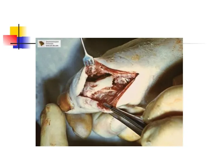

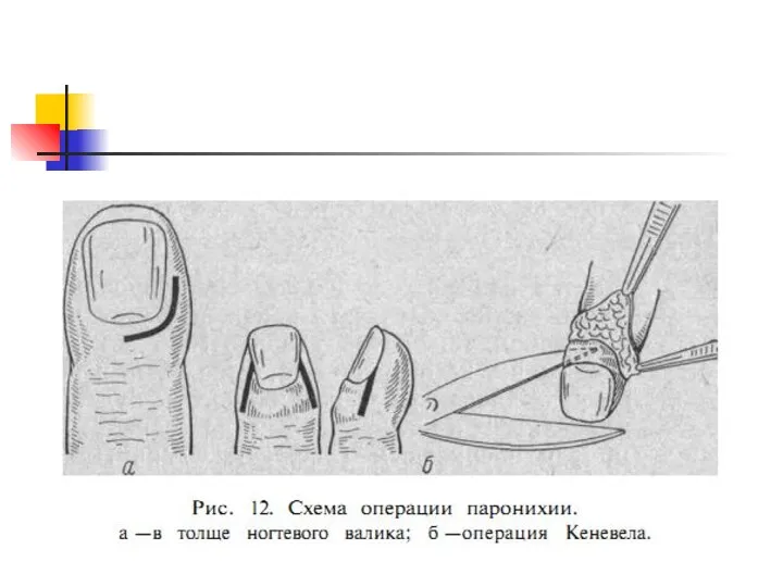
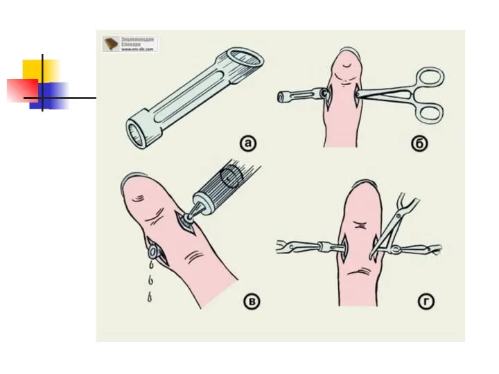
Слайд 27Incisions
Incisions
Слайд 28Phlegmons of the hand
Phlegmons of the hand
Слайд 30forearm
forearm
 Средства для наркоза, снотворные средства. Противосудорожные средства, анальгетики
 Диагностика и лечение мальформации сосудов головного мозга
 Дыхание при различных условиях. Горная болезнь
 Қант диабеті(ҚД) ІІ типі қосарланған созылмалы жүрек жетіспеушілгі (СЖЖ) бар науқастарға стандартты емнің әсері
 Применение ультразвука
 Methadone pharmacogenetics
 Заманауи карпульді анестетиктер. Түрлері. Клиника - фармакологиялық мінездеме
 Классический массаж
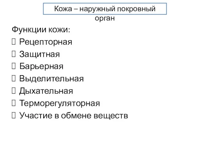 Кожа – наружный покровный орган
 Собрание сообщества психотерапевтов и психологов Оренбургской области
 Причины травматизма в старшем школьном возрасте и пути их предотвращения
 Гастриты. Язвенная болезнь 12 перстной кишки
 Гепатит
 Рандомизированное клиническое исследование ФРИДА. Слайды для врачей отделения нарушения мозгового кровообращения
 Проведение смывов с предметов и рук. Взятие подногтевого соскоба
 Медико-биологические основы физической культуры. Психофизиологические аспекты адаптации человека. (Лекция 3)
 Как дожить до офтальмолога
 Клиническая анатомия лобной пазухи, лобного кармана по данным рентгеновской компьютерной томографии
 Детский невроз. Механизмы и причины развития
 Наука о женщине. История, проблемы и перспективы
 Физиология беременности
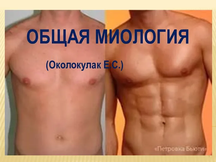 Общая миология
 Требования к обращению с медицинскими отходами
 Болезнь Лайма. Нейроборрелиоз
 Миелопролиферативтік аурулар бір немесе бірнеше гематопоэтических жасушалық желілерін немесе дәнекер тіндік элементтердің
 Сферическая теория артикуляции Монсона
 Физиология-питания
 Диспепсиялық синдром