- Filariasis

Содержание
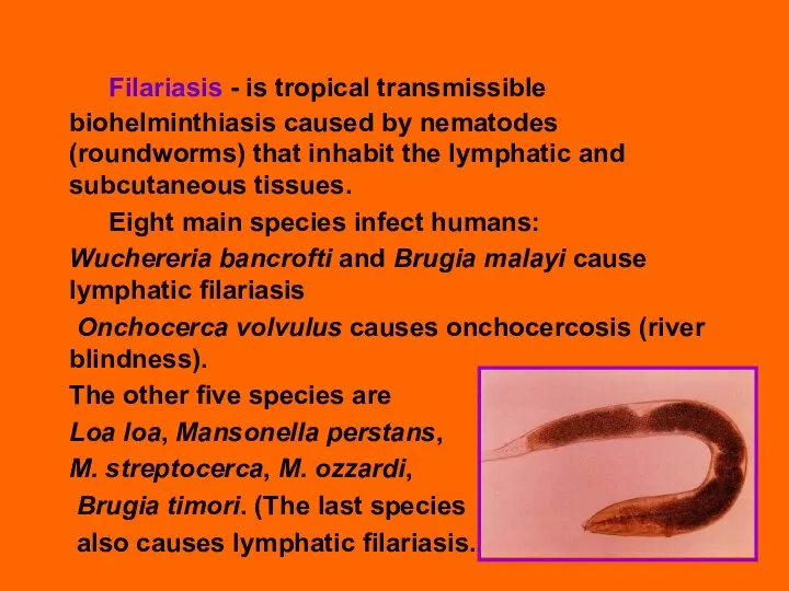
- 2. Filariasis - is tropical transmissible biohelminthiasis caused by nematodes (roundworms) that inhabit the lymphatic and subcutaneous
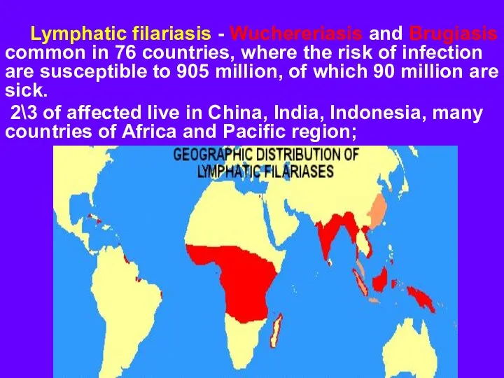
- 3. Lymphatic filariasis - Wuchereriasis and Brugiasis common in 76 countries, where the risk of infection are
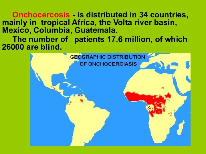
- 4. Onchocercosis - is distributed in 34 countries, mainly in tropical Africa, the Volta river basin, Mexico,
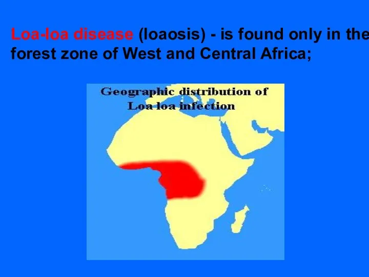
- 5. Loa-loa disease (loaosis) - is found only in the forest zone of West and Central Africa;
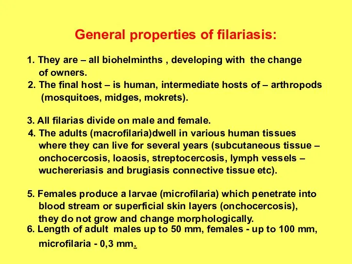
- 6. General properties of filariasis: 1. They are – all biohelminths , developing with the change of
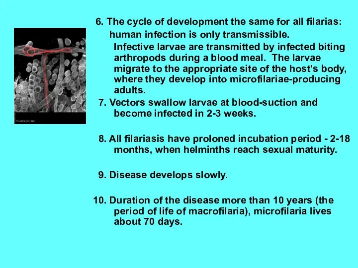
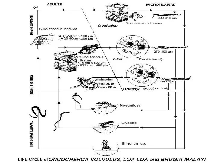
- 7. 6. The cycle of development the same for all filarias: human infection is only transmissible. Infective
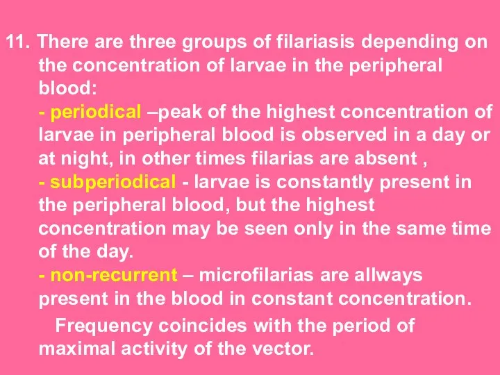
- 9. 11. There are three groups of filariasis depending on the concentration of larvae in the peripheral
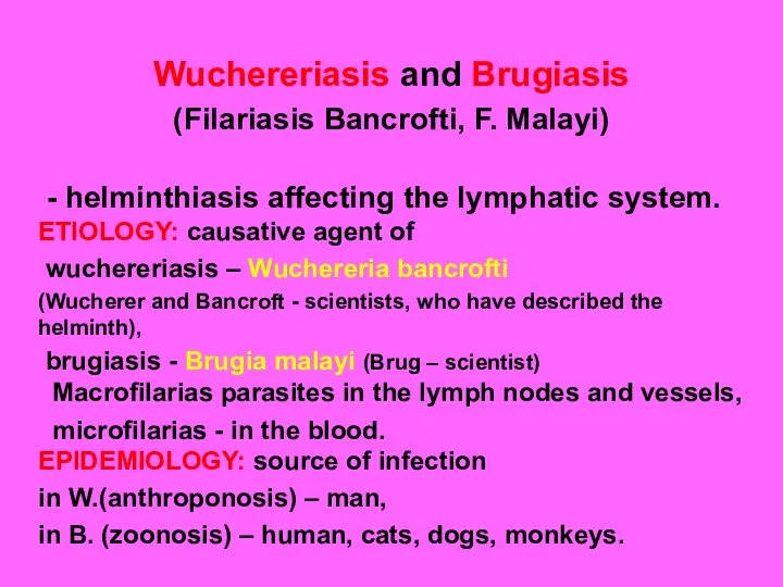
- 10. Wuchereriasis and Brugiasis (Filariasis Bancrofti, F. Malayi) - helminthiasis affecting the lymphatic system. ETIOLOGY: causative agent
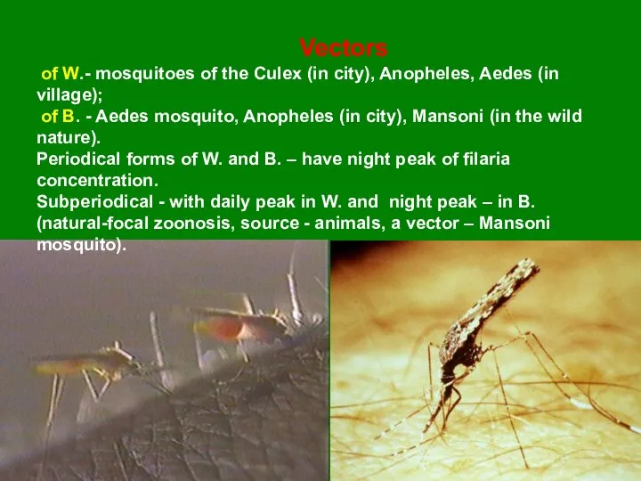
- 11. Vectors of W.- mosquitoes of the Culex (in city), Anopheles, Aedes (in village); of B. -
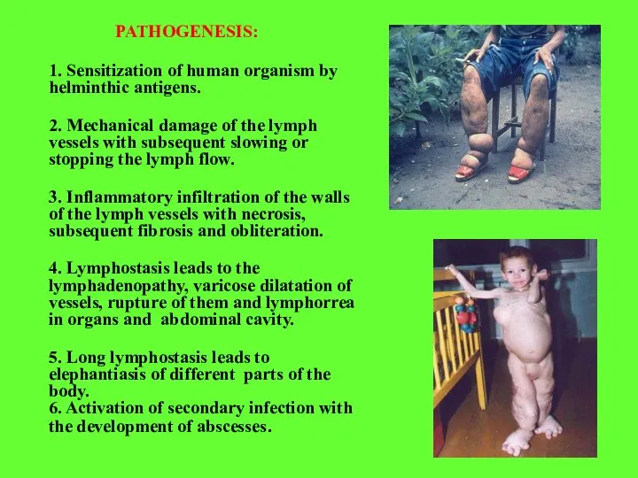
- 12. PATHOGENESIS: 1. Sensitization of human organism by helminthic antigens. 2. Mechanical damage of the lymph vessels
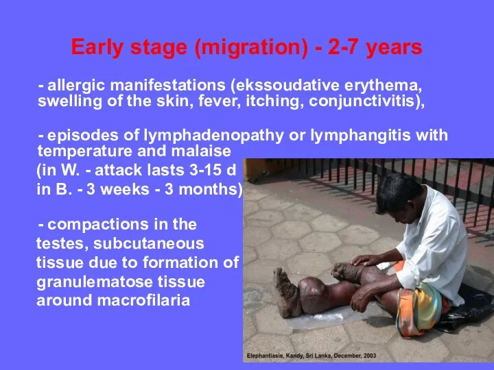
- 13. Early stage (migration) - 2-7 years - allergic manifestations (ekssoudative erythema, swelling of the skin, fever,
- 14. - funikulit, epididymitis, orchitis (in W.) - abscesses in the upper parts of the thighs, under
- 15. rupture of lymph nodes in the kidney, bladder, intestine, mesenterium formation of aseptic abscess around the
- 16. Obstruction stage (develops in 10-15 years): - hydrocele - is the most common manifestation of Wuchereriasis
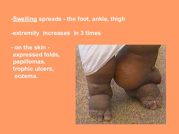
- 18. Swelling spreads - the foot, ankle, thigh extremity increases in 3 times on the skin -
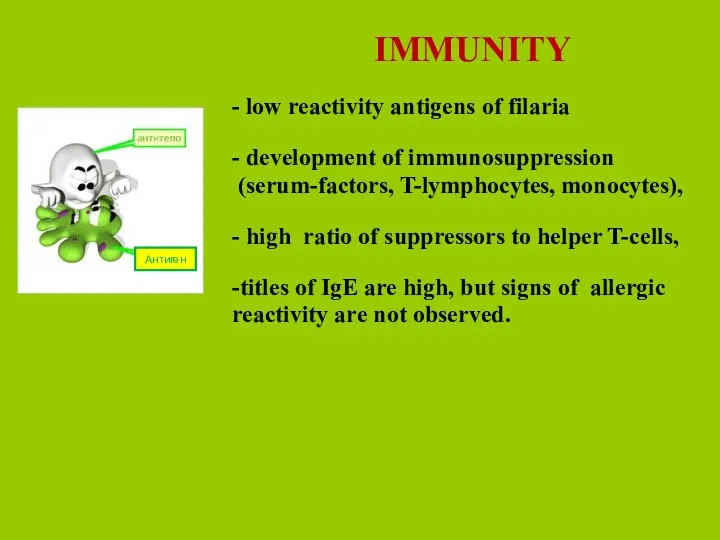
- 19. IMMUNITY - low reactivity antigens of filaria - development of immunosuppression (serum-factors, T-lymphocytes, monocytes), - high
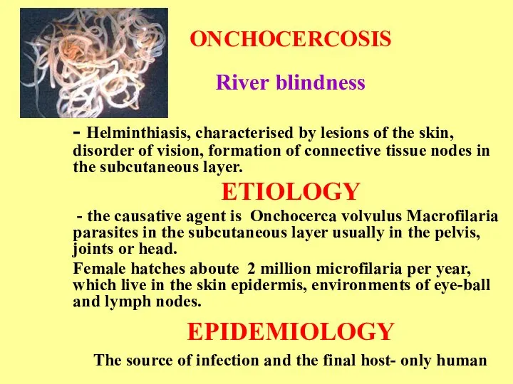
- 20. ONCHOCERCOSIS River blindness - Helminthiasis, characterised by lesions of the skin, disorder of vision, formation of
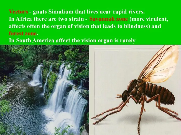
- 21. Vectors - gnats Simulium that lives near rapid rivers. In Africa there are two strain -
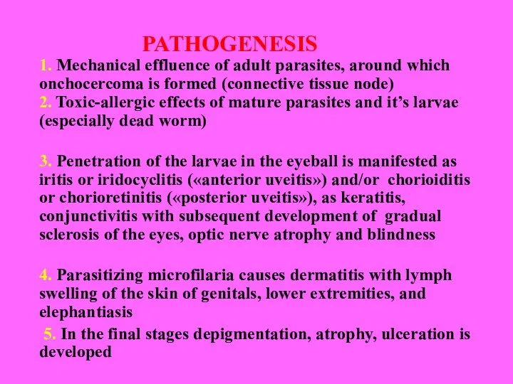
- 22. PATHOGENESIS 1. Mechanical effluence of adult parasites, around which onchocercoma is formed (connective tissue node) 2.
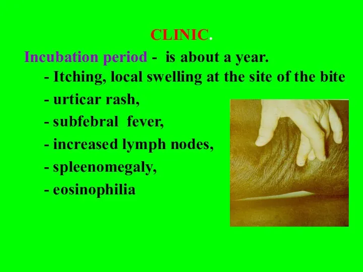
- 23. CLINIC. Incubation period - is about a year. - Itching, local swelling at the site of
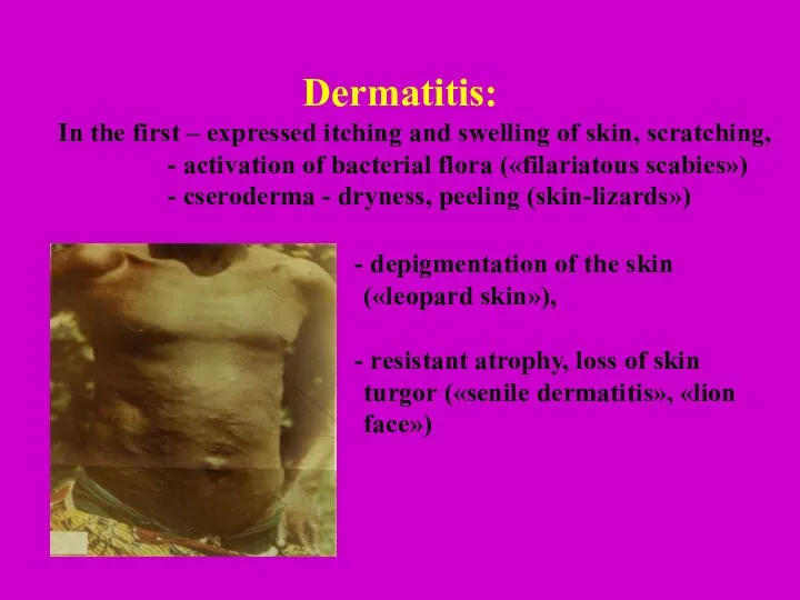
- 24. Dermatitis: In the first – expressed itching and swelling of skin, scratching, - activation of bacterial
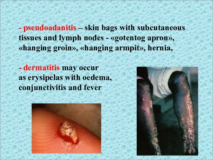
- 25. - pseudoadanitis – skin bags with subcutaneous tissues and lymph nodes - «gotentog apron», «hanging groin»,
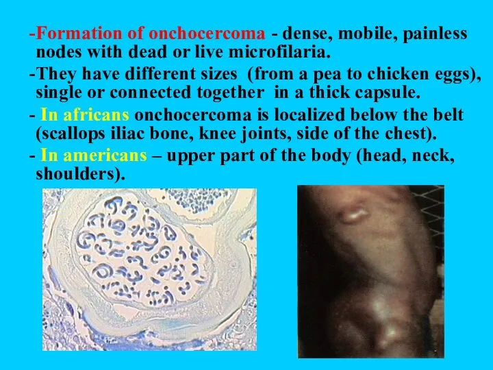
- 26. Formation of onchocercoma - dense, mobile, painless nodes with dead or live microfilaria. They have different
- 27. affection of lymphatic system - lymphadenitis (groin and armpit), lymph oedema,orchitis, hydrocele, elephantiasis of the lower
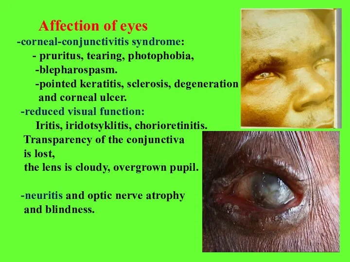
- 28. Affection of eyes corneal-conjunctivitis syndrome: - pruritus, tearing, photophobia, -blepharospasm. -pointed keratitis, sclerosis, degeneration and corneal
- 29. LOAOSIS (Calabar swelling disease) Helminthiasis, characterised by the swelling of soft tissues, affection of eyes and
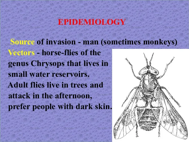
- 30. EPIDEMIOLOGY Source of invasion - man (sometimes monkeys) Vectors - horse-flies of the genus Chrysops that
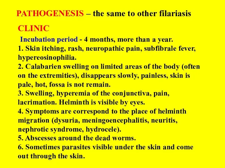
- 31. PATHOGENESIS – the same to other filariasis CLINIC Incubation period - 4 months, more than a
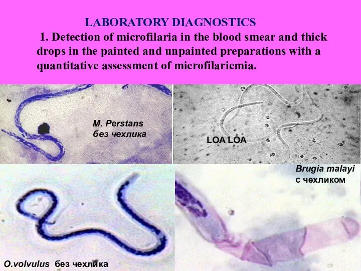
- 32. O.volvulus без чехлика Brugia malayi с чехликом LOA LOA M. Perstans без чехлика LABORATORY DIAGNOSTICS 1.
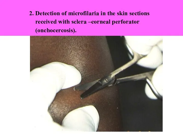
- 33. 2. Detection of microfilaria in the skin sections received with sclera –corneal perforator (onchocercosis).
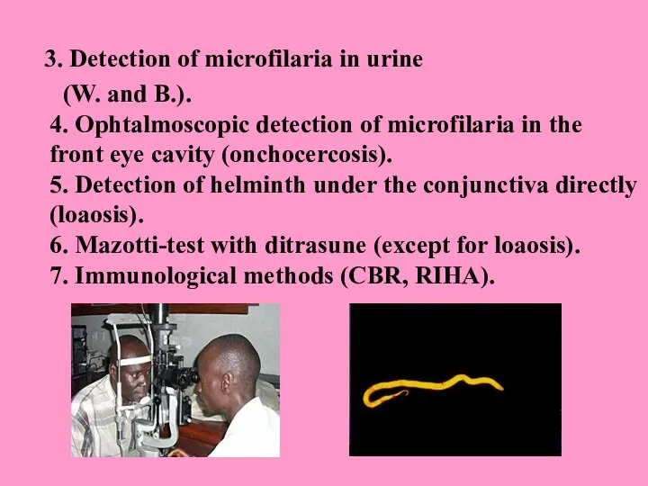
- 34. 3. Detection of microfilaria in urine (W. and B.). 4. Ophtalmoscopic detection of microfilaria in the
- 35. TREATMENT Dietylcarbamasepine - is effective in acute and chronic stage, in latent filariasis 6 mg /kg
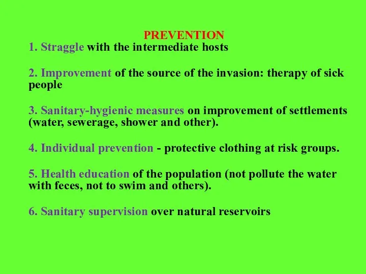
- 36. PREVENTION 1. Straggle with the intermediate hosts 2. Improvement of the source of the invasion: therapy
- 38. Скачать презентацию
Слайд 2 Filariasis - is tropical transmissible biohelminthiasis caused by nematodes (roundworms) that inhabit
Слайд 3
Lymphatic filariasis - Wuchereriasis and Brugiasis common in 76 countries, where the
Lymphatic filariasis - Wuchereriasis and Brugiasis common in 76 countries, where the
Слайд 4 Onchocercosis - is distributed in 34 countries, mainly in tropical Africa, the
Слайд 5Loa-loa disease (loaosis) - is found only in the forest zone of
Loa-loa disease (loaosis) - is found only in the forest zone of
Слайд 6General properties of filariasis:
1. They are – all biohelminths , developing with
1. They are – all biohelminths , developing with
Слайд 7
6. The cycle of development the same for all filarias:
6. The cycle of development the same for all filarias:
Слайд 911. There are three groups of filariasis depending on the concentration of
Слайд 10Wuchereriasis and Brugiasis
(Filariasis Bancrofti, F. Malayi)
- helminthiasis affecting the lymphatic
(Filariasis Bancrofti, F. Malayi)
- helminthiasis affecting the lymphatic
Слайд 11
Vectors
of W.- mosquitoes of the Culex (in city), Anopheles, Aedes (in
Vectors
of W.- mosquitoes of the Culex (in city), Anopheles, Aedes (in
Слайд 12PATHOGENESIS:
1. Sensitization of human organism by helminthic antigens.
2. Mechanical damage of the
1. Sensitization of human organism by helminthic antigens.
2. Mechanical damage of the
Слайд 13Early stage (migration) - 2-7 years
- allergic manifestations (ekssoudative erythema, swelling of
- allergic manifestations (ekssoudative erythema, swelling of
Слайд 14
- funikulit, epididymitis, orchitis (in W.)
- abscesses in the upper
- funikulit, epididymitis, orchitis (in W.)
- abscesses in the upper
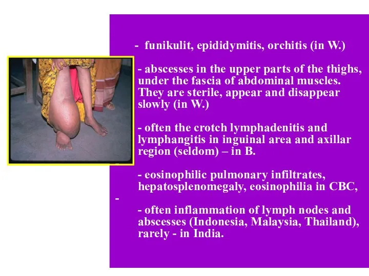
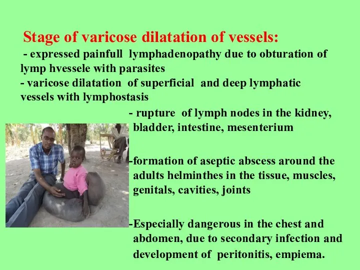
Слайд 15rupture of lymph nodes in the kidney, bladder, intestine, mesenterium
formation of aseptic
formation of aseptic
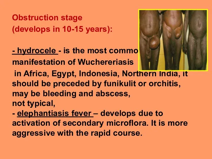
Слайд 16Obstruction stage
(develops in 10-15 years):
- hydrocele - is the most common
(develops in 10-15 years):
- hydrocele - is the most common
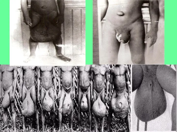
Слайд 18Swelling spreads - the foot, ankle, thigh
extremity increases in 3 times
on
extremity increases in 3 times
on
Слайд 19IMMUNITY
- low reactivity antigens of filaria
- development of immunosuppression
(serum-factors, T-lymphocytes, monocytes),
-
- low reactivity antigens of filaria
- development of immunosuppression
(serum-factors, T-lymphocytes, monocytes),
-
Слайд 20ONCHOCERCOSIS
River blindness
- Helminthiasis, characterised by lesions of the skin, disorder of vision,
- Helminthiasis, characterised by lesions of the skin, disorder of vision,
Слайд 21Vectors - gnats Simulium that lives near rapid rivers.
In Africa there
Vectors - gnats Simulium that lives near rapid rivers.
In Africa there
Слайд 22PATHOGENESIS
1. Mechanical effluence of adult parasites, around which onchocercoma is formed (connective
Слайд 23CLINIC.
Incubation period - is about a year.
- Itching, local
Incubation period - is about a year. - Itching, local
Слайд 24
Dermatitis:
In the first – expressed itching and swelling of skin, scratching,
Dermatitis:
In the first – expressed itching and swelling of skin, scratching,
Слайд 25- pseudoadanitis – skin bags with subcutaneous tissues and lymph nodes -
Слайд 26Formation of onchocercoma - dense, mobile, painless nodes with dead or live
Слайд 27affection of lymphatic system - lymphadenitis (groin and armpit), lymph oedema,orchitis, hydrocele,
affection of lymphatic system - lymphadenitis (groin and armpit), lymph oedema,orchitis, hydrocele,
Слайд 28Affection of eyes
corneal-conjunctivitis syndrome:
- pruritus, tearing, photophobia,
-blepharospasm.
-pointed keratitis,
corneal-conjunctivitis syndrome:
- pruritus, tearing, photophobia,
-blepharospasm.
-pointed keratitis,
Слайд 29LOAOSIS (Calabar swelling disease)
Helminthiasis, characterised by the swelling of soft tissues, affection
Helminthiasis, characterised by the swelling of soft tissues, affection
Слайд 30EPIDEMIOLOGY
Source of invasion - man (sometimes monkeys)
Vectors - horse-flies of
Source of invasion - man (sometimes monkeys)
Vectors - horse-flies of
Слайд 31PATHOGENESIS – the same to other filariasis
CLINIC
Incubation period - 4 months,
PATHOGENESIS – the same to other filariasis
CLINIC
Incubation period - 4 months,
Слайд 32O.volvulus без чехлика
Brugia malayi
с чехликом
LOA LOA
M. Perstans
без чехлика
O.volvulus без чехлика
Brugia malayi
с чехликом
LOA LOA
M. Perstans
без чехлика
Слайд 33
2. Detection of microfilaria in the skin sections
received
2. Detection of microfilaria in the skin sections
received
Слайд 343. Detection of microfilaria in urine
(W. and B.).
4. Ophtalmoscopic detection of
(W. and B.). 4. Ophtalmoscopic detection of
Слайд 35TREATMENT
Dietylcarbamasepine - is effective in acute and chronic stage, in latent filariasis

Слайд 36PREVENTION
1. Straggle with the intermediate hosts
2. Improvement of the source of the
2. Improvement of the source of the
 Алгоритм диагностики и лечения синдрома раздраженного кишечника
 Аллергия. Микробиология
 Общие нарушения в организме при гемобластозах
 Gnano: ощути вкус нанотехнологий. - презентация_
 Медицинская этика и деонтология в медицине
 Методы обследования беременных. Диагностика беременности
 Проблемы современной неонатологии
 Профилактика возникновения профессиональных заболеваний
 Лучевая диагностика опорнодвигательного аппарата
 Оценка эффективности физических методов реабилитации пациентов с острыми нарушениями мозгового кровообращения
 Итоги работы консультанта по вопросам медицинского обеспечения сотрудников компании ООО ПСК ОмскДизель
 Хирургический инструментарий
 Гематология
 Доказанная инволюция фиброза печени
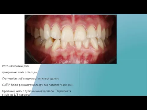 Брекет-система
 Аудиометриялық көрсеткіштері
 ستون فقرات
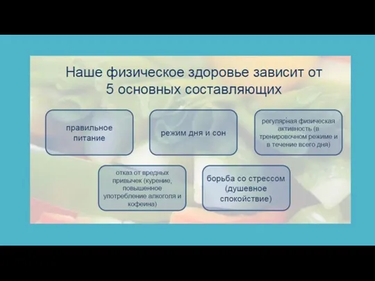 Рекомендации по питанию
 Профилактика меланомы
 Внутричерепные гематомы
 Острый аппендицит Острый панкреатит Тлеубаева Б
 Лечение дислипидемии. СРС 2
 Патогенез приступа стабильной стенокардии
 Влияние воды на действие лекарственных веществ
 I заседание МНК онкологии
 Минеральные вещества
 Артериальное кровотечение. Маточное кровотечение
 Химический состав антибиотиков и альтернативных средств против мастита крупного рогатого скота