- Peptic Ulcer Disease

Содержание
- 2. INTRODUCTION Peptic Ulcer is a lesion in the lining (mucosa) of the digestive tract, typically in
- 3. Lesion may subsequently occur into the lamina propria and submucosa to cause bleeding. – Most of
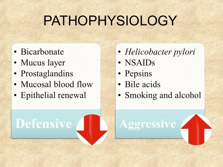
- 4. PATHOPHYSIOLOGY
- 5. Under normal conditions, a physiologic balance exists between gastric acid secretion and gastroduodenal mucosal defense. Mucosal
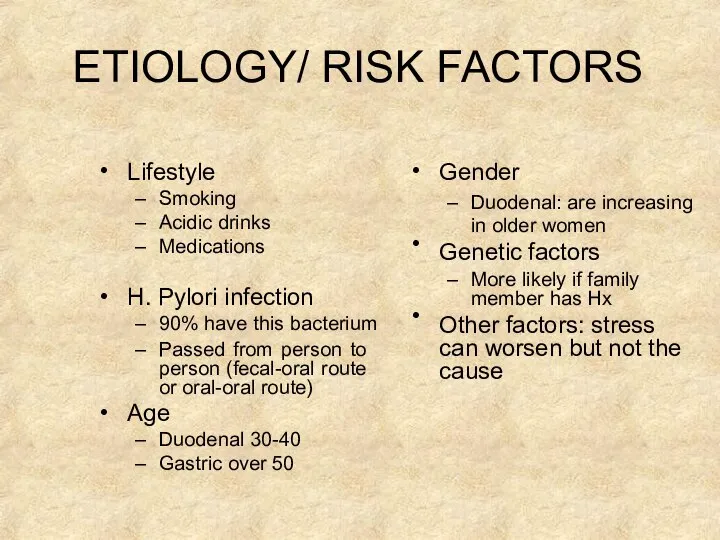
- 6. ETIOLOGY/ RISK FACTORS • Lifestyle Smoking Acidic drinks Medications • • H. Pylori infection 90% have
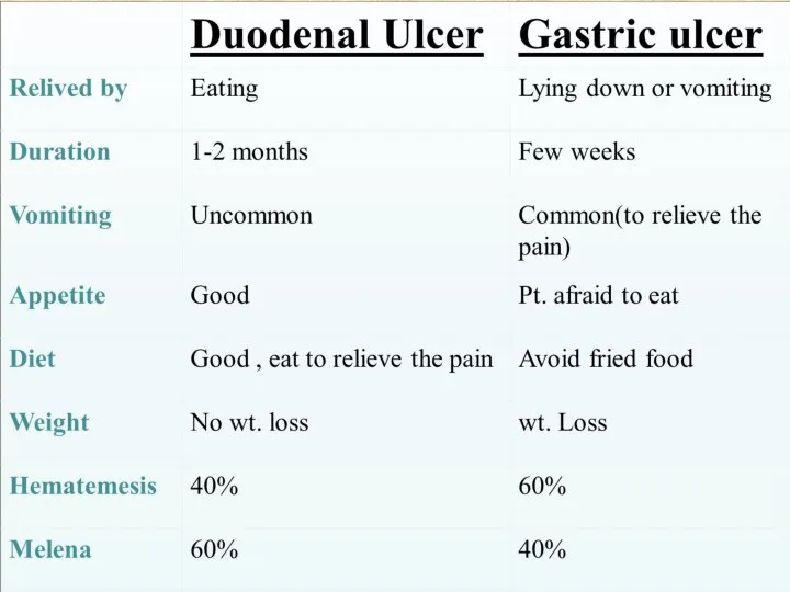
- 7. TYPES GASTRIC PEPTIC ULCER DUODENAL PEPTIC ULCER
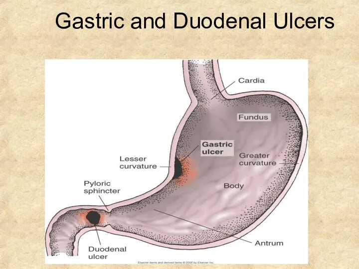
- 8. Gastric and Duodenal Ulcers
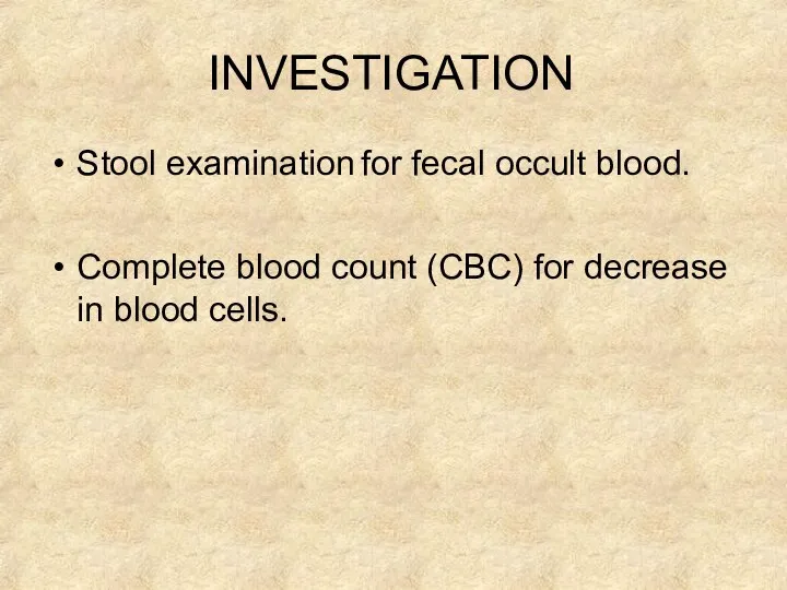
- 11. INVESTIGATION Stool examination for fecal occult blood. Complete blood count (CBC) for decrease in blood cells.
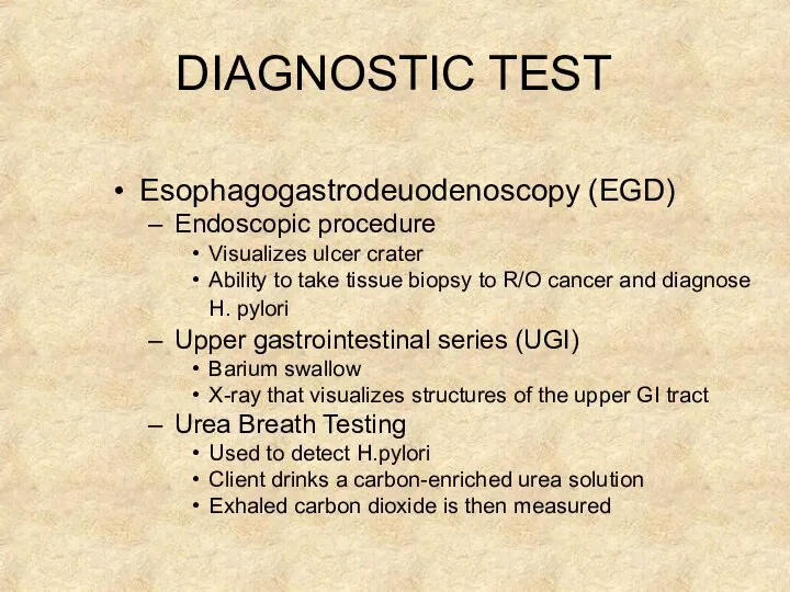
- 12. DIAGNOSTIC TEST Esophagogastrodeuodenoscopy (EGD) Endoscopic procedure Visualizes ulcer crater Ability to take tissue biopsy to R/O
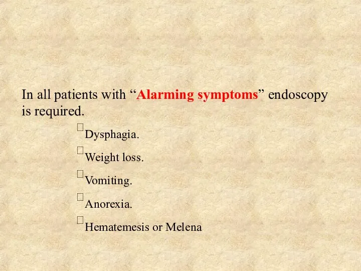
- 13. In all patients with “Alarming symptoms” endoscopy is required. Dysphagia. Weight loss. Vomiting. Anorexia. Hematemesis or
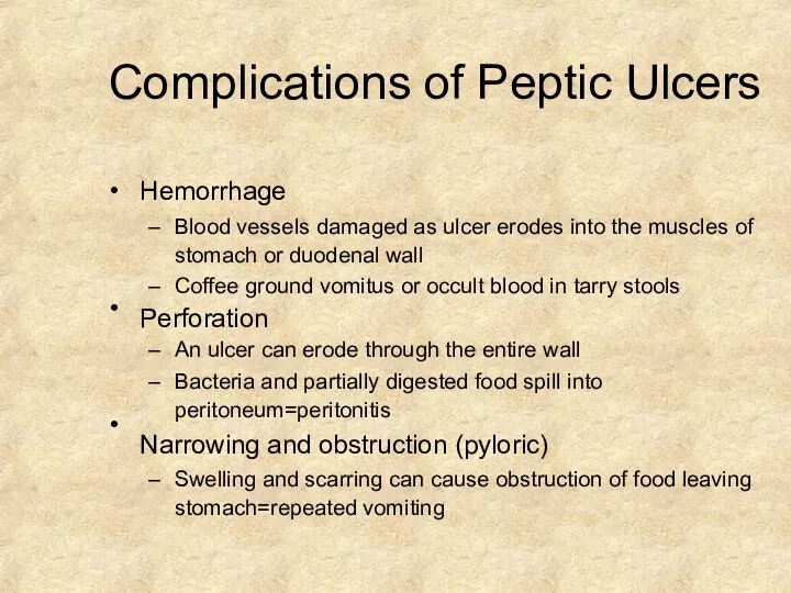
- 14. Complications of Peptic Ulcers • • • Hemorrhage Blood vessels damaged as ulcer erodes into the
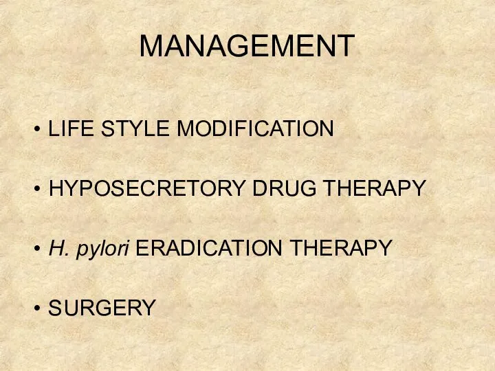
- 15. MANAGEMENT LIFE STYLE MODIFICATION HYPOSECRETORY DRUG THERAPY H. pylori ERADICATION THERAPY SURGERY
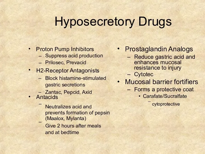
- 17. Hyposecretory Drugs • Proton Pump Inhibitors – – Suppress acid production Prilosec, Prevacid • H2-Receptor Antagonists
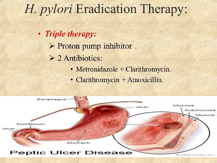
- 18. H. pylori Eradication Therapy:
- 19. Indications: Failure of medical treatment. Development of complications High level of gastric secretion and combined duodenal
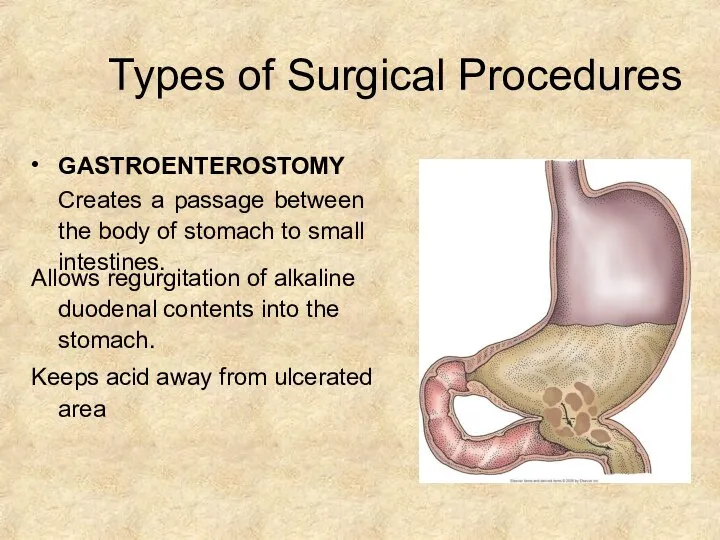
- 20. Types of Surgical Procedures • GASTROENTEROSTOMY Creates a passage between the body of stomach to small
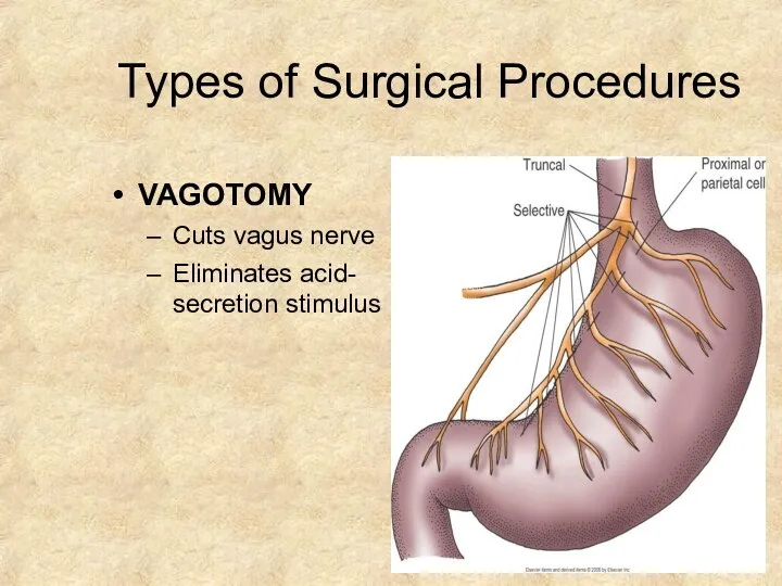
- 21. Types of Surgical Procedures VAGOTOMY Cuts vagus nerve Eliminates acid- secretion stimulus
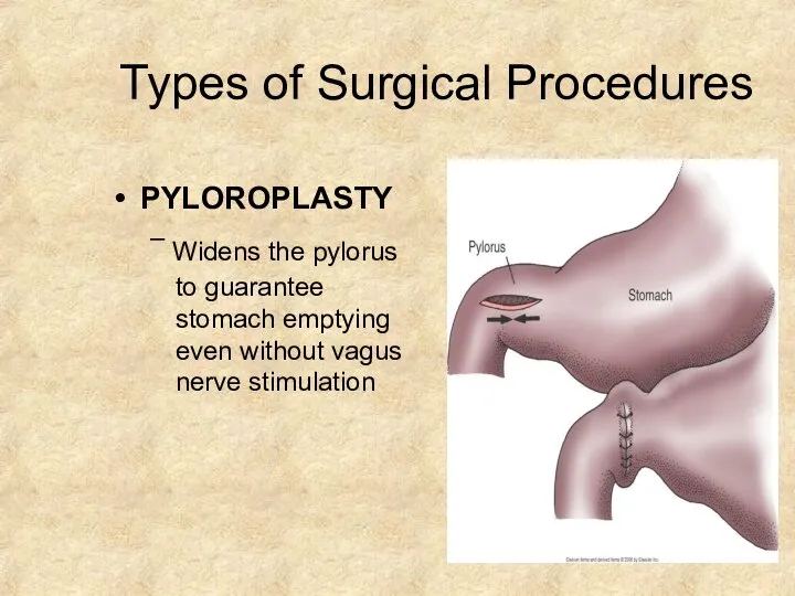
- 22. Types of Surgical Procedures PYLOROPLASTY – Widens the pylorus to guarantee stomach emptying even without vagus
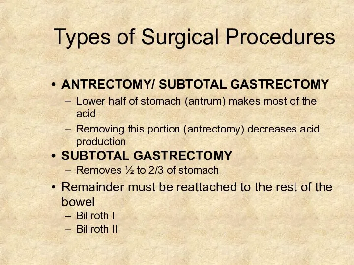
- 23. Types of Surgical Procedures ANTRECTOMY/ SUBTOTAL GASTRECTOMY Lower half of stomach (antrum) makes most of the
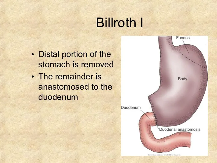
- 24. Billroth I Distal portion of the stomach is removed The remainder is anastomosed to the duodenum
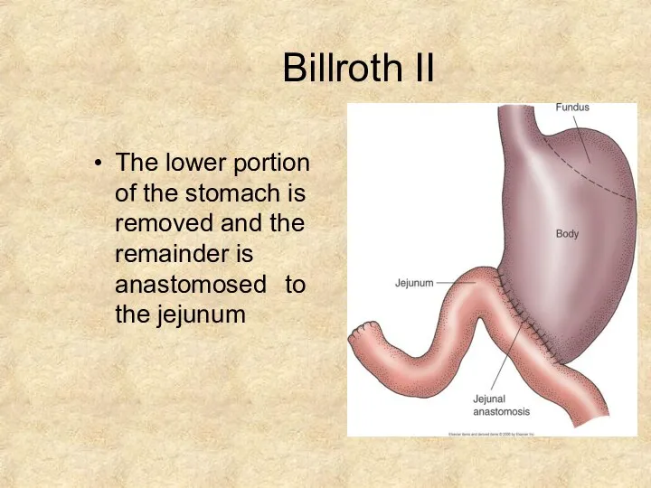
- 25. Billroth II The lower portion of the stomach is removed and the remainder is anastomosed to
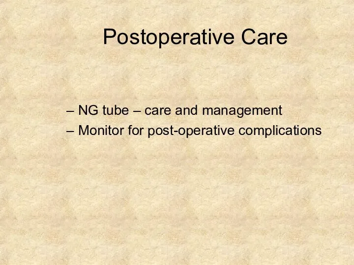
- 26. Postoperative Care NG tube – care and management Monitor for post-operative complications
- 28. Скачать презентацию
Слайд 2INTRODUCTION
Peptic Ulcer is a lesion in the lining
(mucosa) of the digestive tract, typically in the stomach or duodenum, caused by the
digestive action of pepsin and stomach acid.
INTRODUCTION
Peptic Ulcer is a lesion in the lining
(mucosa) of the digestive tract, typically in the stomach or duodenum, caused by the
digestive action of pepsin and stomach acid.
Слайд 3Lesion may subsequently occur into the lamina
propria and submucosa to cause bleeding.
Lesion may subsequently occur into the lamina
propria and submucosa to cause bleeding.
Слайд 4PATHOPHYSIOLOGY
PATHOPHYSIOLOGY
Слайд 5Under normal conditions, a physiologic balance exists between gastric acid secretion and
Under normal conditions, a physiologic balance exists between gastric acid secretion and
Слайд 6ETIOLOGY/ RISK FACTORS
•
Lifestyle
Smoking
Acidic drinks
Medications
•
•
H. Pylori infection
90% have this bacterium
Passed from person to
ETIOLOGY/ RISK FACTORS
•
Lifestyle
Smoking
Acidic drinks
Medications
•
•
H. Pylori infection
90% have this bacterium
Passed from person to
Слайд 7TYPES
GASTRIC PEPTIC ULCER
DUODENAL PEPTIC ULCER
TYPES
GASTRIC PEPTIC ULCER
DUODENAL PEPTIC ULCER
Слайд 8Gastric and Duodenal Ulcers
Gastric and Duodenal Ulcers
Слайд 11INVESTIGATION
Stool examination for fecal occult blood.
Complete blood count (CBC) for decrease in blood
INVESTIGATION
Stool examination for fecal occult blood.
Complete blood count (CBC) for decrease in blood
Слайд 12DIAGNOSTIC TEST
Esophagogastrodeuodenoscopy (EGD)
Endoscopic procedure
Visualizes ulcer crater
Ability to take tissue biopsy to R/O
DIAGNOSTIC TEST
Esophagogastrodeuodenoscopy (EGD)
Endoscopic procedure
Visualizes ulcer crater
Ability to take tissue biopsy to R/O
Слайд 13In all patients with “Alarming symptoms” endoscopy is required.
Dysphagia.
Weight loss.
Vomiting.
Anorexia.
Hematemesis or Melena
In all patients with “Alarming symptoms” endoscopy is required.
Dysphagia.
Weight loss.
Vomiting.
Anorexia.
Hematemesis or Melena
Слайд 14Complications of Peptic Ulcers
•
•
•
Hemorrhage
Blood vessels damaged as ulcer erodes into the muscles of
Complications of Peptic Ulcers
•
•
•
Hemorrhage
Blood vessels damaged as ulcer erodes into the muscles of
Слайд 15MANAGEMENT
LIFE STYLE MODIFICATION
HYPOSECRETORY DRUG THERAPY
H. pylori ERADICATION THERAPY
SURGERY
MANAGEMENT
LIFE STYLE MODIFICATION
HYPOSECRETORY DRUG THERAPY
H. pylori ERADICATION THERAPY
SURGERY
Слайд 17Hyposecretory Drugs
•
Proton Pump Inhibitors
–
–
Suppress acid production Prilosec, Prevacid
•
H2-Receptor Antagonists
Block histamine-stimulated gastric secretions
Zantac,
Hyposecretory Drugs
•
Proton Pump Inhibitors
–
–
Suppress acid production Prilosec, Prevacid
•
H2-Receptor Antagonists
Block histamine-stimulated gastric secretions
Zantac,
Слайд 18H. pylori Eradication Therapy:
H. pylori Eradication Therapy:
Слайд 19Indications:
Failure of medical treatment.
Development of complications
High level of gastric secretion and combined
Indications:
Failure of medical treatment.
Development of complications
High level of gastric secretion and combined
Слайд 20Types of Surgical Procedures
•
GASTROENTEROSTOMY
Creates a passage between the body of stomach to
Types of Surgical Procedures
•
GASTROENTEROSTOMY
Creates a passage between the body of stomach to
Слайд 21Types of Surgical Procedures
VAGOTOMY
Cuts vagus nerve
Eliminates acid- secretion stimulus
Types of Surgical Procedures
VAGOTOMY
Cuts vagus nerve
Eliminates acid- secretion stimulus
Слайд 22Types of Surgical Procedures
PYLOROPLASTY
– Widens the pylorus to guarantee stomach emptying even
Types of Surgical Procedures
PYLOROPLASTY
– Widens the pylorus to guarantee stomach emptying even
Слайд 23Types of Surgical Procedures
ANTRECTOMY/ SUBTOTAL GASTRECTOMY
Lower half of stomach (antrum) makes most
Types of Surgical Procedures
ANTRECTOMY/ SUBTOTAL GASTRECTOMY
Lower half of stomach (antrum) makes most
Слайд 24Billroth I
Distal portion of the stomach is removed
The remainder is anastomosed to
Billroth I
Distal portion of the stomach is removed
The remainder is anastomosed to
Слайд 25Billroth II
The lower portion of the stomach is removed and the remainder
Billroth II
The lower portion of the stomach is removed and the remainder
Слайд 26Postoperative Care
NG tube – care and management
Monitor for post-operative complications
Postoperative Care
NG tube – care and management
Monitor for post-operative complications
Похожие презентации
 Плечевое сплетение, симптомы тотального поражения
 Влияние психотропных средств на кроветворение
 Инструменты реализации решений Правительства РФ по противодействию ВИЧ/СПИД
 Реализация дифференцированного подхода в формировании артикуляционного праксиса у дошкольников с артикуляционными расстройствами
 Внелёгочный туберкулёз
 Неотложные состояния в гастроэнтерологии
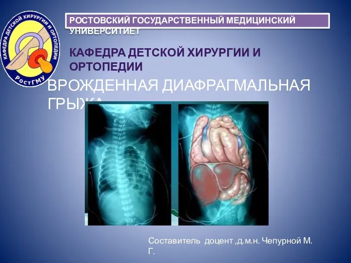 Врожденная диафрагмальная грыжа
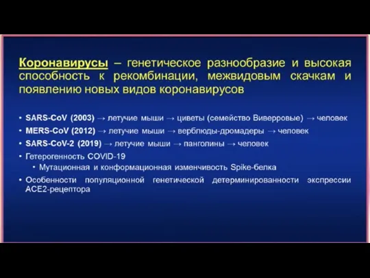 Коронавирус
 Схемы реализации методов. Метод сенсорной интеграции. Метод Бобат. Методика Перфетти
 Болезни почек
 Іш қуысы ағзаларын ультрадыбыстық зерттеу
 Распил таза
 ТУР-синдром
 Тест. Клиникалық жағдай
 Острый лимфобластный лейкоз
 Клиническая картина повреждения прямой кишки
 Патобиохимический аспект развития первичного гемохроматоза и его клинических симптомов
 Көпір тәрізді протездің тарихы, маңыздылығы, мақсаты
 Гликогеновая болезнь
 Оптическая система глаза
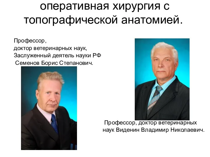 Оперативная хирургия с топографической анатомией
 Опыт использования ООО Автолан-Плюс программно-аппаратного комплекса Телемедик
 Правила здорового питания детей
 Урологиядағы лабораторлы зерртеу әдістері
 Нейрореаниматология – современные аспекты
 Местная анестезия
 Микробиологические аспекты