- Recidivance Leishmaniasis (Lupoid)

Содержание
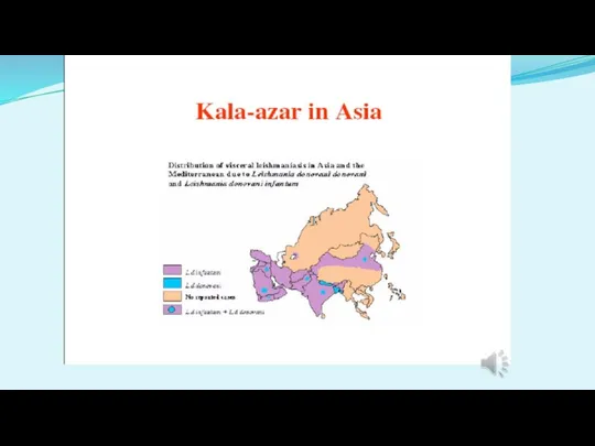
- 2. Old World Visceral Leishmaniasis (OWVL)
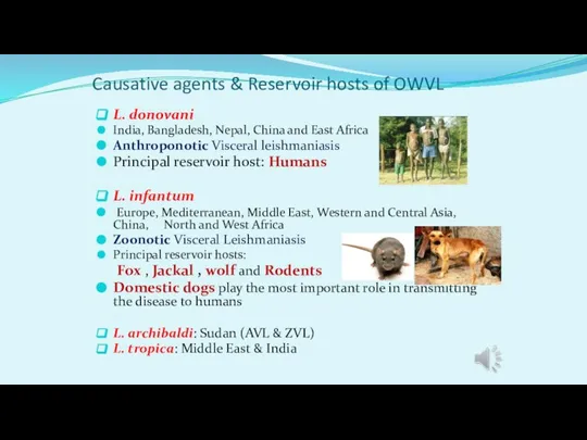
- 3. Causative agents & Reservoir hosts of OWVL L. donovani India, Bangladesh, Nepal, China and East Africa
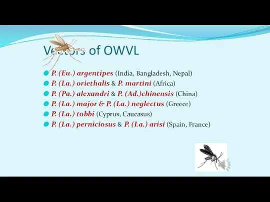
- 4. Vectors of OWVL P. (Eu.) argentipes (India, Bangladesh, Nepal) P. (La.) oriethalis & P. martini (Africa)
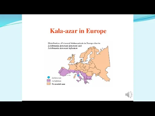
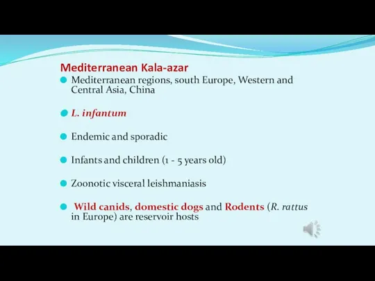
- 9. Mediterranean Kala-azar Mediterranean regions, south Europe, Western and Central Asia, China L. infantum Endemic and sporadic
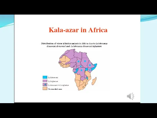
- 10. African Kala-azar L. donovani: AVL , East Africa L. infantum: ZVL , North and West Africa
- 11. African grass Rat
- 12. Indian Kala-azar India, Bangladesh, Nepal, East Africa, China L. donovani Humans are principal reservoir hosts Adults
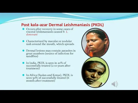
- 13. Post kala-azar Dermal Leishmaniasis (PKDL) Occurs after recovery in some cases of visceral leishmaniasis caused b
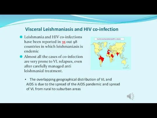
- 14. Visceral Leishmaniasis and HIV co-infection Leishmania and HIV co-infections have been reported in 35 out 98
- 15. Clinical symptoms in VL in Human Prolonged Irregular fever Hepatomegaly Splenomegali Anemia , Leukopenia and Thrombocytopenia
- 16. Clinical symptoms in VL in Human Splenomegaly, distended abdomen and severe muscle wasting.
- 17. Clinical symptoms in VL in Human Splenomegaly , severe muscle wasting and Cachexia
- 18. Clinical symptoms in VL in Human Hepatomegaly and Splenomegali in an autopsy of an infant dying
- 19. Clinical symptoms in VL in Human Jaundice hands of a VL patient
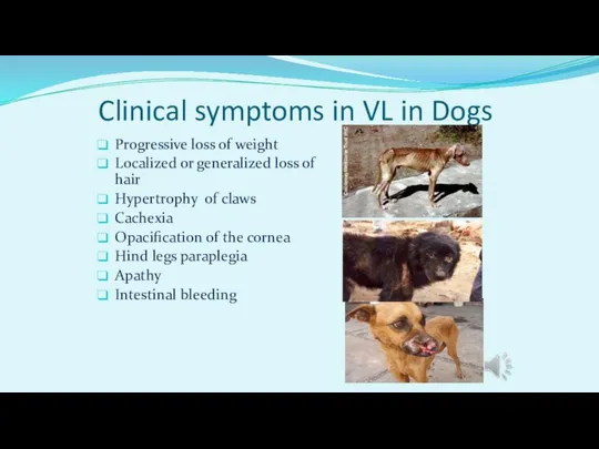
- 20. Clinical symptoms in VL in Dogs Progressive loss of weight Localized or generalized loss of hair
- 21. Diagnosis of VL The incubation period for VL is 10 days to 1 year (or 2
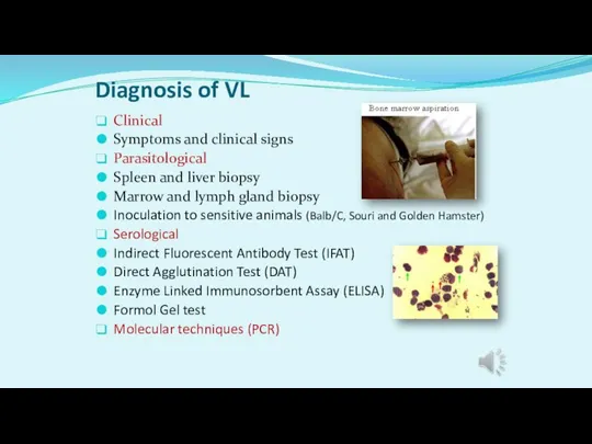
- 22. Diagnosis of VL Clinical Symptoms and clinical signs Parasitological Spleen and liver biopsy Marrow and lymph
- 24. Скачать презентацию
Слайд 3Causative agents & Reservoir hosts of OWVL
L. donovani
India, Bangladesh, Nepal, China
Causative agents & Reservoir hosts of OWVL
L. donovani
India, Bangladesh, Nepal, China
Слайд 4Vectors of OWVL
P. (Eu.) argentipes (India, Bangladesh, Nepal)
P. (La.) oriethalis & P.
Vectors of OWVL
P. (Eu.) argentipes (India, Bangladesh, Nepal)
P. (La.) oriethalis & P.
Слайд 9Mediterranean Kala-azar
Mediterranean regions, south Europe, Western and Central Asia, China
L. infantum
Endemic and
Mediterranean Kala-azar
Mediterranean regions, south Europe, Western and Central Asia, China
L. infantum
Endemic and
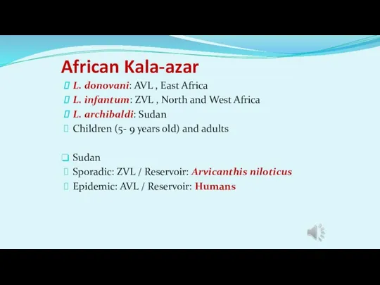
Слайд 10African Kala-azar
L. donovani: AVL , East Africa
L. infantum: ZVL , North and
African Kala-azar
L. donovani: AVL , East Africa
L. infantum: ZVL , North and
Слайд 11African grass Rat
African grass Rat

Слайд 12Indian Kala-azar
India, Bangladesh, Nepal, East Africa, China
L. donovani
Humans are principal reservoir hosts
Adults
Indian Kala-azar
India, Bangladesh, Nepal, East Africa, China
L. donovani
Humans are principal reservoir hosts
Adults
Слайд 13Post kala-azar Dermal Leishmaniasis (PKDL)
Occurs after recovery in some cases of visceral
Post kala-azar Dermal Leishmaniasis (PKDL)
Occurs after recovery in some cases of visceral
Слайд 14Visceral Leishmaniasis and HIV co-infection
Leishmania and HIV co-infections have been reported in
Visceral Leishmaniasis and HIV co-infection
Leishmania and HIV co-infections have been reported in
Слайд 15Clinical symptoms in VL in Human
Prolonged Irregular fever
Hepatomegaly
Splenomegali
Anemia , Leukopenia and Thrombocytopenia
Clinical symptoms in VL in Human
Prolonged Irregular fever
Hepatomegaly
Splenomegali
Anemia , Leukopenia and Thrombocytopenia

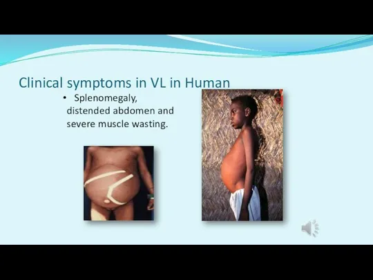
Слайд 16Clinical symptoms in VL in Human
Splenomegaly, distended abdomen and severe muscle
Clinical symptoms in VL in Human
Splenomegaly, distended abdomen and severe muscle
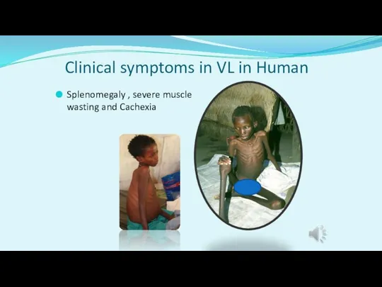
Слайд 17Clinical symptoms in VL in Human
Splenomegaly , severe muscle wasting and Cachexia
Clinical symptoms in VL in Human
Splenomegaly , severe muscle wasting and Cachexia
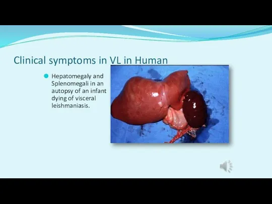
Слайд 18Clinical symptoms in VL in Human
Hepatomegaly and Splenomegali in an autopsy of
Clinical symptoms in VL in Human
Hepatomegaly and Splenomegali in an autopsy of
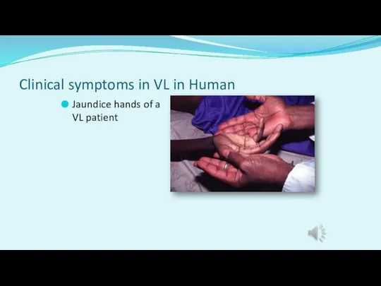
Слайд 19Clinical symptoms in VL in Human
Jaundice hands of a VL patient
Clinical symptoms in VL in Human
Jaundice hands of a VL patient
Слайд 20Clinical symptoms in VL in Dogs
Progressive loss of weight
Localized or generalized
Clinical symptoms in VL in Dogs
Progressive loss of weight
Localized or generalized
Слайд 21Diagnosis of VL
The incubation period for VL is 10 days to 1
Diagnosis of VL
The incubation period for VL is 10 days to 1
Слайд 22Diagnosis of VL
Clinical
Symptoms and clinical signs
Parasitological
Spleen and liver biopsy
Marrow and lymph gland
Diagnosis of VL
Clinical
Symptoms and clinical signs
Parasitological
Spleen and liver biopsy
Marrow and lymph gland
 Ультразвуковая диагностика в гинекологии
 Денсаулыққа байланысты өмір сапасы
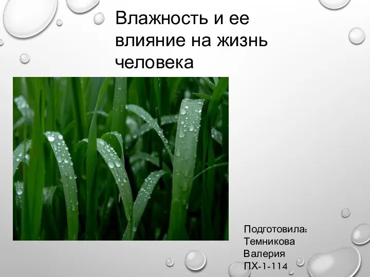 Влажность и ее влияние на жизнь человека
 Гранулематоз с полиангиитом (Гранулематоз Вегенера)
 Жедел контрацепция
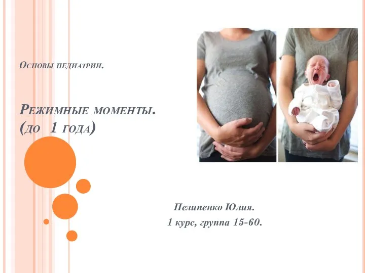 Основы педиатрии. Режимные моменты (дети до 1 года)
 Топография нижней конечности
 Детская гемиплегия
 Неонатальный скрининг
 Пневмоперитонеум. Виды пневмоперитонеума
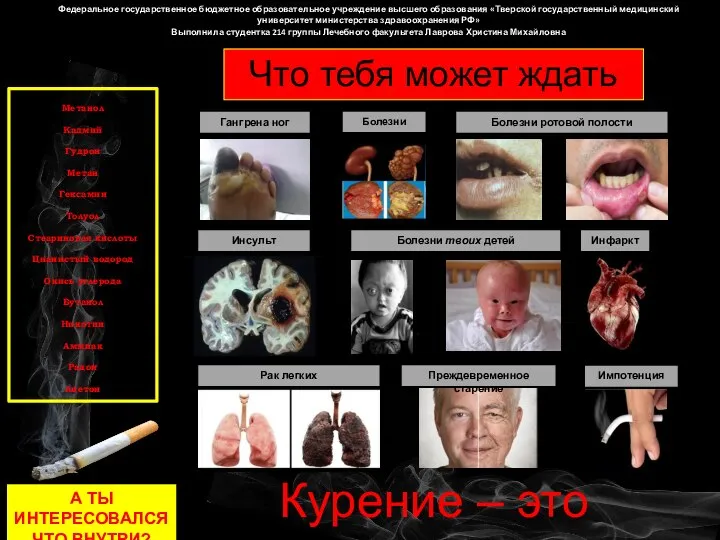 Патологии курения
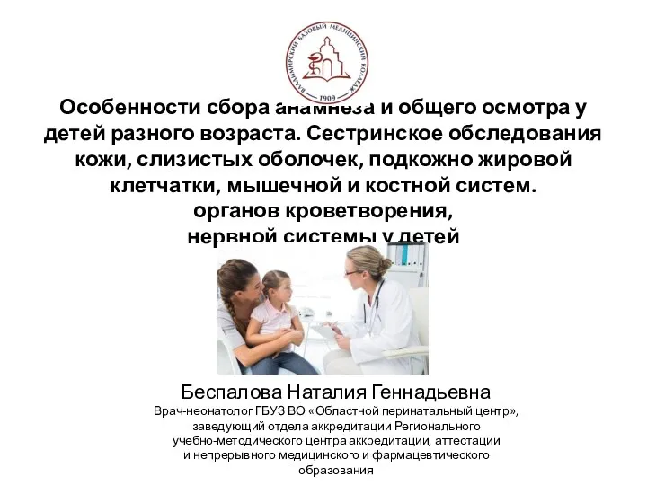 Тема 1 Анамнез.Сестринское обследование кожи и костно-мышечной системы
 Социально-этические проблемы генетики
 Роль ГБО в лечении и профилактике преэклампсии
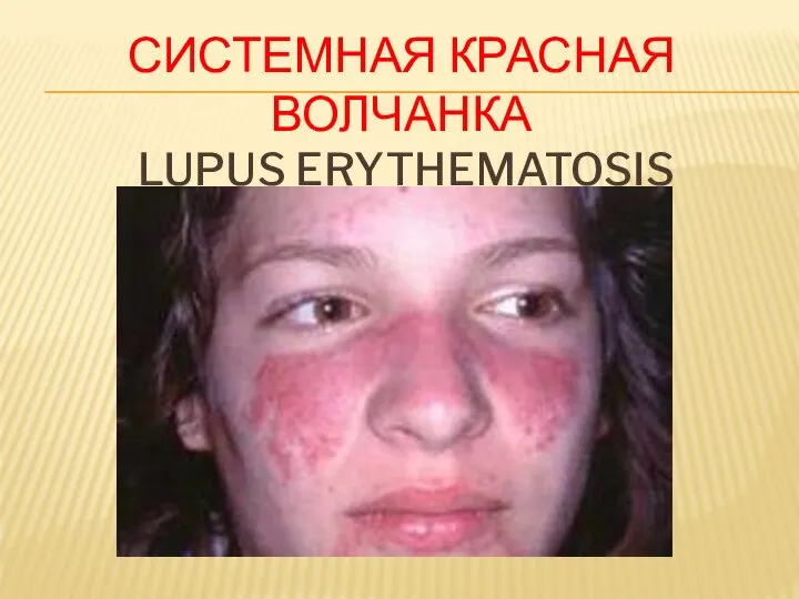 Системная красная волчанка
 Связки и сухожилия. Опорно-двигательный аппарат
 Первичные иммунодефициты (ПИД)
 Показатели развития отрасли здравоохранения Российской Федерации за 2012 – 2017 гг
 Kontingenční tabulky - SAS
 Инфекционный мононуклеоз
 Реабилитация при пояснично-крестцовом остеохондрозе
 Евгеника. Евгеника түрлері. Евгеника және қазіргі заман
 Средства, влияющие на афферентную иннервацию
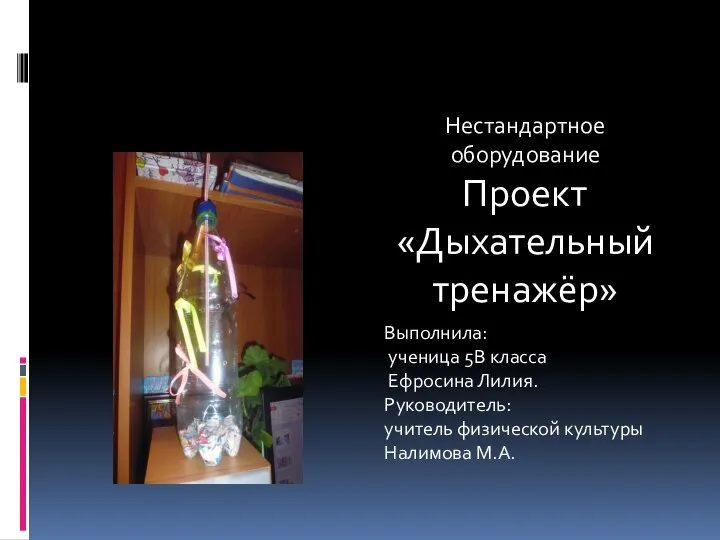 Дыхательный тренажёр
 Наномедицина. Наномедицинаның қолдану аясы
 Этиология, патогенез и классификация гемобластозов
 Изготовление мостовидных конструкций в ортопедической стоматологии
 Қантты диабет. Диабеттік фетопатия