- Results of treatment of vascular tumors of head

Содержание
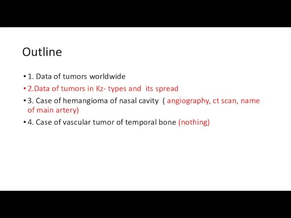
- 2. Outline 1. Data of tumors worldwide 2.Data of tumors in Kz- types and its spread 3.
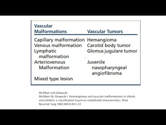
- 3. Mulliken and Glowacki Mulliken JB, Glowacki J. Hemangiomas and vascular malformations in infants and children: a
- 4. Paragangliomas account for 0.6% of all neoplasms in the head and neck region and 0.03% of
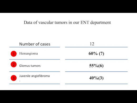
- 5. Data of vascular tumors in our ENT department 1 2 3
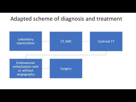
- 6. Adapted scheme of diagnosis and treatment
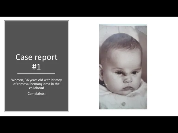
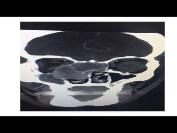
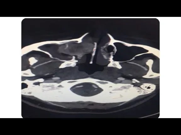
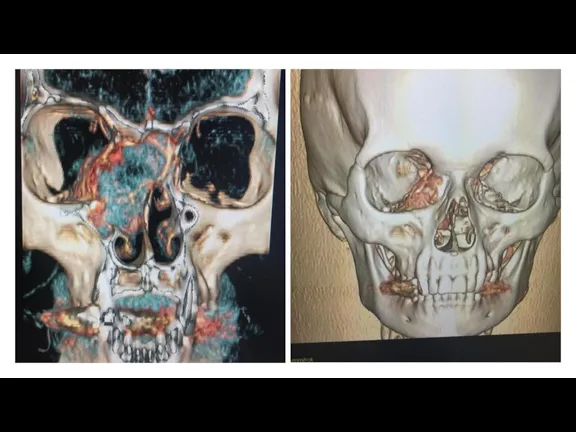
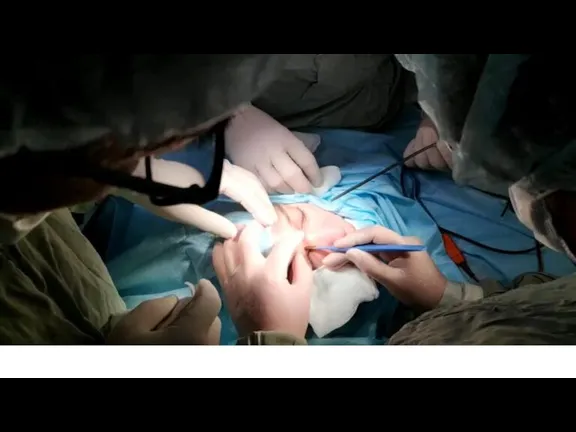
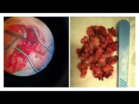
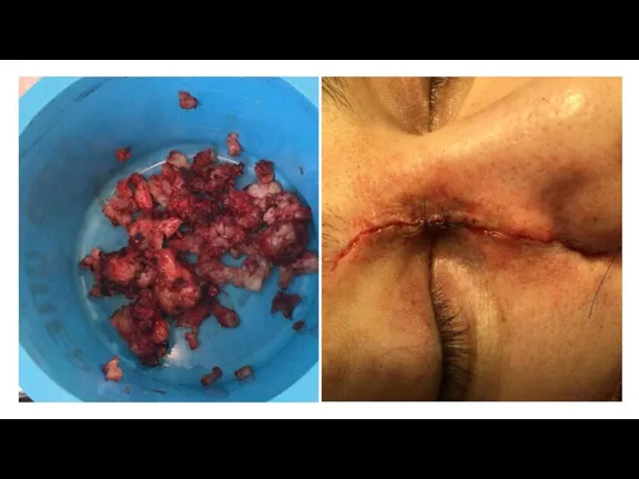
- 7. Case report #1 Women, 36 years old with history of removal hemangioma in the childhood Complaints:
- 14. Case report #2: glomus tumor of temporal bone
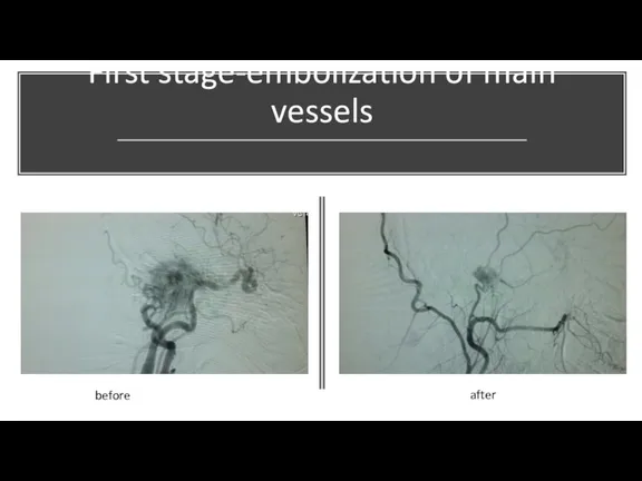
- 15. First stage-embolization of main vessels before after
- 18. Conclusions: 1. Using modern technologies such as contrast CT scan allowed to visualize the borders of
- 20. Скачать презентацию
Слайд 3Mulliken and Glowacki
Mulliken JB, Glowacki J. Hemangiomas and vascular malformations in
Mulliken and Glowacki
Mulliken JB, Glowacki J. Hemangiomas and vascular malformations in
Слайд 4Paragangliomas account for 0.6% of all neoplasms in the head and neck
Paragangliomas account for 0.6% of all neoplasms in the head and neck
Слайд 5Data of vascular tumors in our ENT department
1
2
3
Data of vascular tumors in our ENT department
1
2
3
Слайд 6Adapted scheme of diagnosis and treatment
Adapted scheme of diagnosis and treatment
Слайд 7Case report #1
Women, 36 years old with history of removal hemangioma in
Case report #1
Women, 36 years old with history of removal hemangioma in
Слайд 14Case report #2: glomus tumor of temporal bone
Case report #2: glomus tumor of temporal bone
Слайд 15First stage-embolization of main vessels
before
after
First stage-embolization of main vessels
before
after
Слайд 18Conclusions:
1. Using modern technologies such as contrast CT scan allowed to visualize
Conclusions:
1. Using modern technologies such as contrast CT scan allowed to visualize
 Опухоли носа и околоносовых пазух
 Заболевания печени. Современный взгляд на лечение и профилактику
 Анатомия и физиология женской репродуктивной системы
 УЗИ органов брюшной полости
 Мы живем в мире, где есть СПИД
 Заболевания гипофиза (Акромегалия, гипофизарный нанизм, пролактинома, несахарный диабет, гормональнонеактивные опухоли гипофиза)
 Инструменты реализации решений Правительства РФ по противодействию ВИЧ/СПИД
 Лекарственные средства, влияющие на афферентную инервацию
 Фармакопрофилактика және фармакотерапия туралы түсінік. Фармакотерапия түрлері. Рационалды фармакотерапия принциптері
 Туберкулинадиагностика
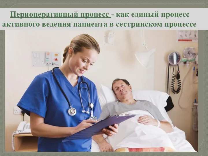 Периоперативный процесс как единый процесс активного ведения пациента в сестринском процессе
 Свертывание крови. Группы крови. Переливание. (8 класс)
 Профилактика инфекционных заболеваний
 The immediate (early phase) allergic reaction in the nose
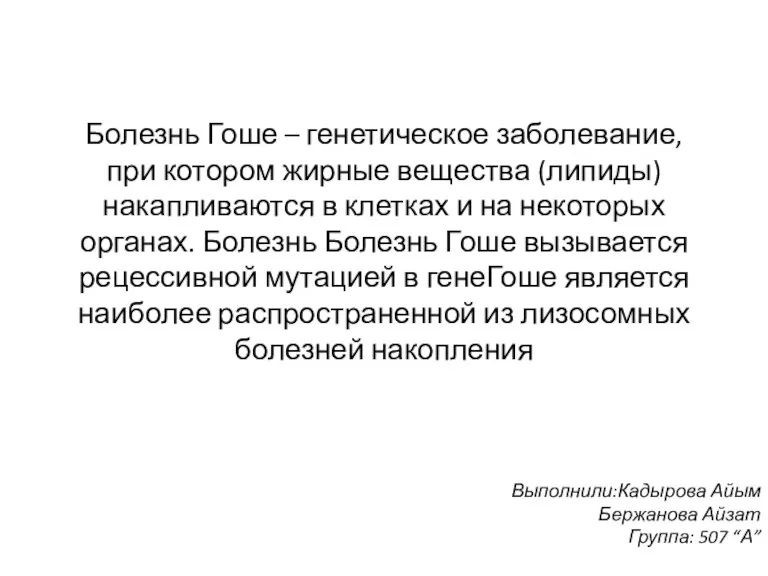 Болезнь Гоше
 Грипп и ОРВИ
 Острая массивная кровопотеря (ОМК)
 Грипп и вакцинопрофилактика
 История болезни. Острый нелимфобластный лейкоз
 Suturas periodontales
 Первая помощь при острой сердечной недостаточности и инсульте
 Острый аппендицит
 Рентгенография желудка и двенадцатиперстной кишки с двойным контрастированием
 Ультразвук. Применение в науке
 Добавочный скелет. Соединение костей
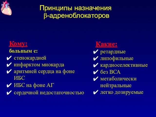 Принципы назначения бета-адреноблокаторов
 Жировые эмульсии
 Состояния СОПР при заболеваниях внутренних органов и систем. Травматические поражения СОПР